. 2019 Feb 13:23:5.
doi: 10.1186/s40824-019-0154-1. eCollection 2019.

Preservation of allograft bone using a glycerol solution: a compilation of original preclinical research

Affiliations

Preservation of allograft bone using a glycerol solution: a compilation of original preclinical research

Brian Samsell et al. Biomater Res. .

Abstract

Background: Bone allografts are used in many orthopedic procedures to provide structural stability as well as an osteoconductive matrix for bone ingrowth and fusion. Traditionally, bone allografts have been preserved by either freezing or freeze-drying. Each of these preservation methods has some disadvantages: Frozen grafts require special shipping and storage conditions, and freeze-drying requires special lyophilization equipment and procedures that may impact biomechanical integrity. This report describes an alternate type of preservation using glycerol, which allows storage of fully-hydrated tissues at ambient temperature avoiding the potential complications from freeze-drying.

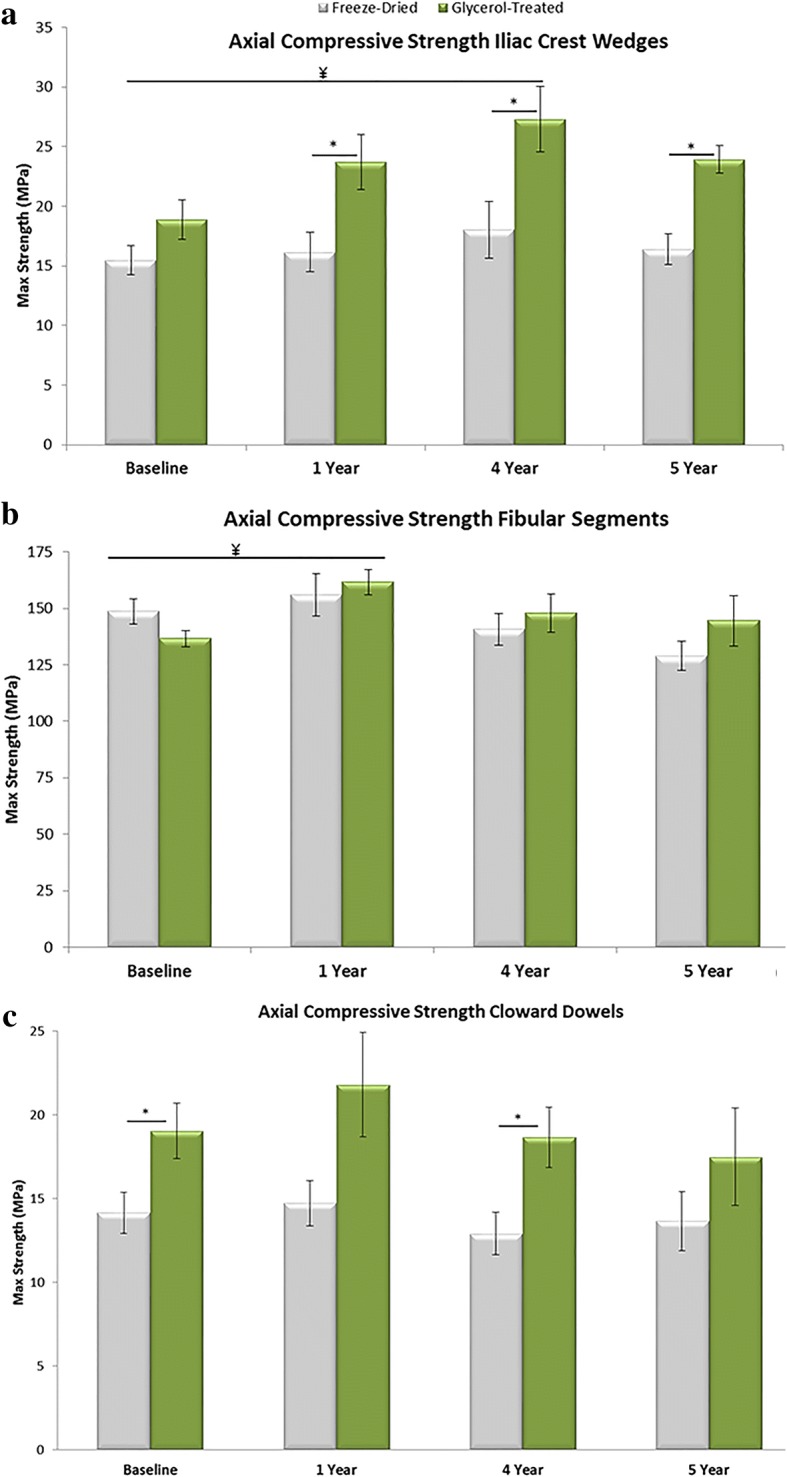
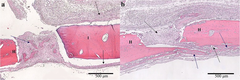
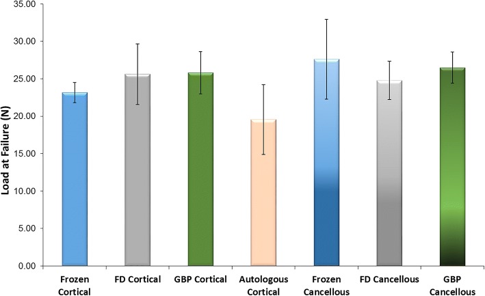
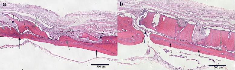
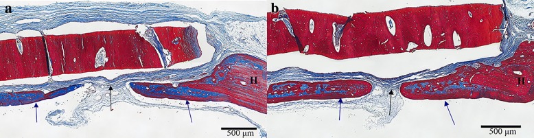
Methods: In the in vitro three-point bend test, cortical bone was processed and frozen, freeze-dried, or treated with glycerol-based preservation (GBP). Load was applied to each graft at a rate of 2.71 mm/min. The flexural strain, flexural strength, and flexural modulus were then calculated. In the in vitro axial compression test, iliac crest wedges, fibular segments, and Cloward dowels were processed and either freeze-dried or GBP treated. The compressive strength of the grafts were tested at time zero and after real time aging of 1, 4, and 5 years. In the in vivo rat calvarial defect assessment, freeze-dried, frozen, and GBP bone implants were compared after being implanted into a critical sized defect. Samples underwent histological and biomechanical evaluation.

Results: Bone grafts subjected to GBP were found to be at least biomechanically equivalent to frozen bone while also being significantly less brittle than freeze-dried bone. GBP-preserved bone demonstrated significantly greater compressive strength than freeze-dried at multiple time points. Preclinical research performed in calvaric defect models found that GBP-preserved bone had similar osteoconductivity and biocompatibility to frozen and freeze-dried samples.

Conclusion: Preclinical research demonstrated that glycerol-preservation of bone yields a material that maintains biomechanical strength while eliminating the need for extensive rehydration or thaw periods if used clinically. Additionally, in vivo evidence suggests no negative impact of glycerol-preservation on the ability of bone grafts to successfully participate in new bone formation and fusion.

Keywords: Allograft; Freeze-dried; Frozen; Glycerol; Preservon; Tissue preservation.


Conflict of interest statement

The in vivo assessment was approved by the University of Maryland Institutional Animal Care and Use Committee (IACUC) (reference number 04–09-04).Not applicable.BS, DS, XQ, JM, PS, KG, and MM are employees of LifeNet Health, a non-profit organization that invented the glycerol preservation technology described here.Springer Nature remains neutral with regard to jurisdictional claims in published maps and institutional affiliations.

Figures

Fig. 1
Fig. 1
Examples of bone that have undergone glycerol-perseveration (GBP): a ilium strip with two cortical sides and a cancellous matrix, b cancellous bone block, and c cancellous bone dowel with thin cortical plate. Reproduced with permission from LifeNet Health
Fig. 2
Fig. 2
Flexural strain and flexural strength of the three preservation groups as evaluated using a three-point bend test. a Average maximum flexural strain. The glycerol-preservation (GBP) and frozen groups were not significantly different, but both demonstrated significantly greater average maximum flexural strain compared to the freeze-dried group. b Average maximum flexural strength. The GBP, frozen, and freeze-dried groups were each significantly different from one another
Fig. 3
Fig. 3
Axial compressive strength of different bone grafts treated with glycerol-preservation (GBP) or freeze-drying a) Axial compressive strength of iliac crest wedges. b Axial compressive strength of fibular segments. c Axial compressive strength of Cloward dowels. ¥ statistical significance between GBP at a given time point and both baseline groups (p < 0.05). *statistical significance between GBP and freeze-dried groups at given time point (p < 0.05)
Fig. 4
Fig. 4
a Glycerol-preserved (GBP) cortical bone disc in calvaric defect model at 1 week. The scale is 500 μm. Black arrows indicate the connective tissue surrounding the implant, which resembles normal fibroblast-like cell organization. The blue arrow emphasizes new bone development in the host-implant junction (blue arrow). b Autogenous bone disc in calvaric defect model at 1 week. The scale is 500 μm. Black arrows indicate connective tissue infiltration, and blue arrows mark new bone development initiating the bone bridge formation. *I = Implant bone. H = Host bone
Fig. 5
Fig. 5
Average load at failure for rat calvarial defect – deformation analysis. Glycerol-preservation (GBP) groups were not statistically different than freeze-dried groups for any parameter (p > 0.05)
Fig. 6
Fig. 6
a Glycerol-preserved (GBP) cancellous bone in a tight-fit rat calvarial defect. The scale is 500 μm. b Freeze-dried cortical bone in a tight-fit rat calvarial defect at 6 weeks. The scale is 500 μm. Black arrows mark complete bone bridge formation. Blue arrows indicate soft tissue infiltrate. Green arrows mark osteointegration. H = Host bone. I = Implant bone
Fig. 7
Fig. 7
a Glycerol-preserved (GBP) cortical bone disc in calvaric defect model at 6 weeks. The scale is 500 μm. b Frozen bone disc in calvaric defect model at 6 weeks. The scale is 500 μm. For both images, black arrows indicate connective tissue infiltration and blue arrows mark new bone development initiating the bone bridge formation
Fig. 8
Fig. 8
Six months postoperative X-ray images showing fusion for 2-level ACDF treated using frozen allografts (left) and glycerol-preserved (GBP) allografts (right). Reproduced from Rodway and Gander [23] under the Creative Commons Attribution License, which permits unrestricted use, distribution, and reproduction in any medium, provided the original work is properly cited

References

    1. McGuire DA, Hendricks SD. Allograft tissue in ACL reconstruction. Sports Med Arthrosc. 2009;17(4):224–233. doi: 10.1097/JSA.0b013e3181c0cf8a. - DOI - PubMed
    1. Buser Z, Brodke DS, Youssef JA, Meisel HJ, Myhre SL, Hashimoto R, et al. Synthetic bone graft versus autograft or allograft for spinal fusion: a systematic review. J Neurosurg Spine. 2016;25(4):509–516. doi: 10.3171/2016.1.SPINE151005. - DOI - PubMed
    1. Han JH, Kim HJ, Song JG, Yang JH, Bhandare NN, Fernandez AR, et al. Is bone grafting necessary in opening wedge high Tibial osteotomy? A meta-analysis of radiological outcomes. Knee Surg Relat Res. 2015;27(4):207–220. doi: 10.5792/ksrr.2015.27.4.207. - DOI - PMC - PubMed
    1. Monje A, Pikos MA, Chan HL, Suarez F, Gargallo-Albiol J, Hernandez-Alfaro F, et al. On the feasibility of utilizing allogeneic bone blocks for atrophic maxillary augmentation. Biomed Res Int. 2014;2014:814578. doi: 10.1155/2014/814578. - DOI - PMC - PubMed
    1. Boyce T, Edwards J, Scarborough N. Allograft bone: the influence of processing on safety and performance. Orthop Clin N Am. 1999;30(4):571–581. doi: 10.1016/S0030-5898(05)70110-3. - DOI - PubMed