. 2019 May 7;47(8):4319-4333.
doi: 10.1093/nar/gkz125.

Structure, dynamics and roX2-lncRNA binding of tandem double-stranded RNA binding domains dsRBD1,2 of Drosophila helicase Maleless

Affiliations

Structure, dynamics and roX2-lncRNA binding of tandem double-stranded RNA binding domains dsRBD1,2 of Drosophila helicase Maleless

Pravin Kumar Ankush Jagtap et al. Nucleic Acids Res. .

Abstract

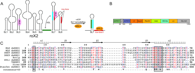
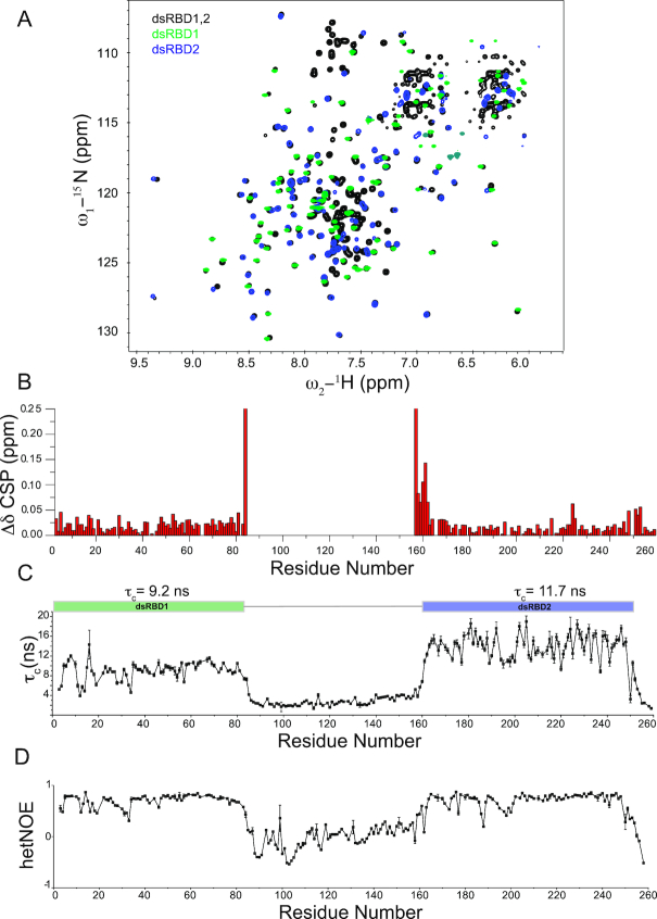
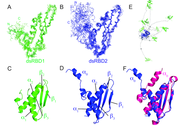
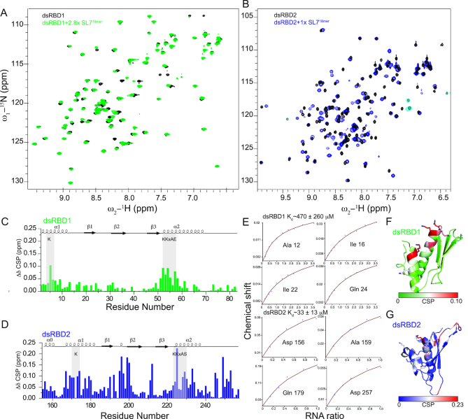
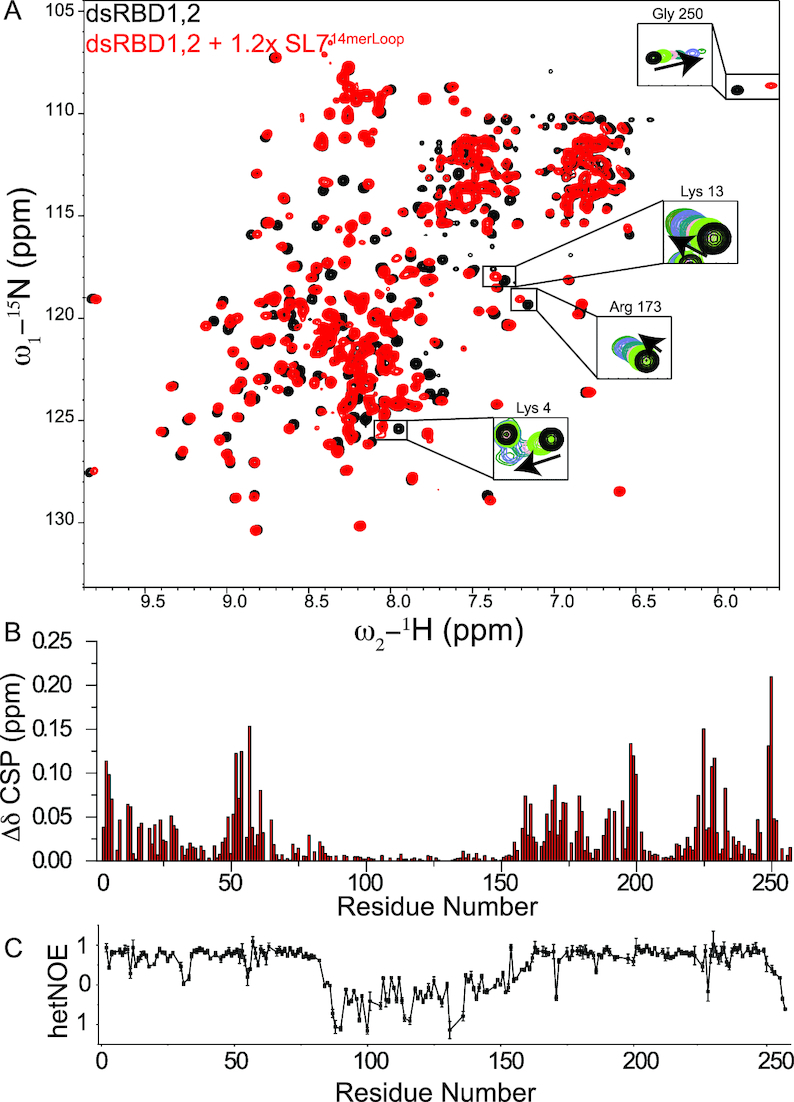
Maleless (MLE) is an evolutionary conserved member of the DExH family of helicases in Drosophila. Besides its function in RNA editing and presumably siRNA processing, MLE is best known for its role in remodelling non-coding roX RNA in the context of X chromosome dosage compensation in male flies. MLE and its human orthologue, DHX9 contain two tandem double-stranded RNA binding domains (dsRBDs) located at the N-terminal region. The two dsRBDs are essential for localization of MLE at the X-territory and it is presumed that this involves binding roX secondary structures. However, for dsRBD1 roX RNA binding has so far not been described. Here, we determined the solution NMR structure of dsRBD1 and dsRBD2 of MLE in tandem and investigated its role in double-stranded RNA (dsRNA) binding. Our NMR and SAXS data show that both dsRBDs act as independent structural modules in solution and are canonical, non-sequence-specific dsRBDs featuring non-canonical KKxAXK RNA binding motifs. NMR titrations combined with filter binding experiments and isothermal titration calorimetry (ITC) document the contribution of dsRBD1 to dsRNA binding in vitro. Curiously, dsRBD1 mutants in which dsRNA binding in vitro is strongly compromised do not affect roX2 RNA binding and MLE localization in cells. These data suggest alternative functions for dsRBD1 in vivo.


Figures

Figure 1.
Figure 1.
(A) Secondary structure of roX2 RNA consisting of eight stem loops. Stem-loops SL7 and SL8 contain roX-boxes (shown in red). Upon remodelling by MLE, the intervening linker between SL6 and SL7 (green) can base pair with the nucleotides from SL7 (cyan) to form an alternative stem (ASL), thus creating a binding site for MSL2 (10). (B) Domain arrangement of MLE as derived from the MLE crystal structure. (C) Structure based sequence alignment of dsRBD1 and dsRBD2 from MLE with DHX9 dsRBDs (Homo sapiens, PDB ID: 3VYX and 3VYY), Rnt1p (Saccharomyces cerevisiae, PDB ID: 1T4N), dsRBD2 domain of HYL1 (Arabidopsis thaliana, PDB ID: 2L2M), TAR RNA binding protein 2 (Homo sapiens, PDB ID: 3LLH), DICER (Schizosaccharomyces pombe, PDB ID: 2L6M) and dsRBD3 domain of Staufen (Drosophila melanogaster, PDB ID: 1EKZ). Secondary structure with respect to MLE dsRBD1 is shown on top of the sequence alignment. Consensus sequence with >60% identity is shown at the bottom of the alignment. RNA binding regions in the dsRBD domains are indicated with gray squares.
Figure 2.
Figure 2.
(A) 1H,15N-HSQC NMR spectrum of dsRBD1,2 (black), dsRBD1 (green) and dsRBD2 (blue) are shown. No major chemical shift differences between the three spectra could be observed suggesting that the two domains act as independent modules in solution. (B) Chemical shift perturbations in dsRBD1,2 compared to individual domains. Residues located at the C-terminus of dsRBD1 and the N-terminus of dsRBD2 are the only ones which exhibit chemical shift differences, but this derives from being the terminal residues in case of individual domains and being connected to the linker in context of the dsRBD1,2 construct. (C) Rotational correlation times (τc) for individual residues as calculated from the ratio of 15N R2/R1 relaxation rates are shown. Average τc for each individual domain is shown on top of the graph. Error bars are calculated from duplicate relaxation delays (see methods). (D) {1H}–15N heteronuclear NOE values for the MLE-dsRBD1,2. Both graphs visualize the different dynamics of the linker region compared to the domain regions. The linker region is highly dynamic.
Figure 3.
Figure 3.
NMR ensemble of 10 lowest energy structures generated by backbone superposition of (A) dsRBD1 residue 1–84 and (B) dsRBD2 (residue 147–259) in the MLE-dsRBD1,2 NMR structure. The N- and C-termini of the two domains are marked for clarity. Cartoon representations of (C) dsRBD1 and (D) dsRBD2 are shown along with the labelled secondary structure elements. (E) Superposition of dsRBD2 (blue) backbone in the dsRBD1,2 ensemble shows that dsRBD1 (green) does not adopt any fixed orientation relative to dsRBD1 owing to the lack of NOE’s between the two domains. Linker residues are shown in grey. (F) Comparison of the solution structure of dsRBD2 presented in this work (blue) and the crystal structure of dsRBD2 as part of MLEcore (PDB ID 5AOR) (magenta). The major difference is the orientation of helix α0 with respect to the dsRBD domain. However, our NMR relaxation data and absence of NOEs between α0 and domain residues indicate that in the absence of other MLE domains this helix is dynamic.
Figure 4.
Figure 4.
(A, B) Superposition of 1H, 15N HSQC NMR spectra of individual dsRBD1 and dsRBD2 domains free and bound to SL718mer. Only the final titration point is shown here for clarity (see Supplementary Figure S4 for all titration points). (CD) Chemical shift perturbations upon titration with saturating concentrations of SL718mer in dsRBD1 and dsRBD2 respectively. (E) Fitted NMR titration curves are shown along with the affinity calculated from the average of peaks showing fast exchange. Errors were calculated by error propagation of fitting error for individual peaks used for affinity calculation. (FG) Mapping of chemical shift perturbations on the NMR structure of dsRBD1 and dsRBD2, respectively.
Figure 5.
Figure 5.
(A) Comparison of 1H,15N-HSQC NMR spectra of dsRBD1,2 free (black) and bound to excess concentrations of SL714merLoop (red). Full titration points for some of the residues are shown in the insets and Supplementary Figure S8. (B) Chemical shift perturbations in dsRBD1,2 upon titration with SL714merLoop. (C) {1H}–15N heteronuclear NOE analysis of dsRBD1,2 bound to SL714merLoop. Error bars are calculated from the spectral noise.
Figure 6.
Figure 6.
(A) Filter binding experiments of MLEfl-dsRBD mutants with SL7 RNA. Error bars represent standard deviation of two experiments. (B) Enrichment of GFP-MLE and its mutants on X chromosome territory is shown. Error bars represent standard deviations for two independent biological replicates. (C) Representative images of analyzed cells for quantification of enrichment of GFP-MLE in X chromosome territory is shown. DNA counterstaining with DAPI and immunostaining against GFP, MLE and MSL2 along with the merged image for GFP and MSL2 channel is shown. n1 and n2 are the number of cells analyzed in two independent biological replicates respectively. The white scale bar in the DAPI channel image represents 5 μm.

Similar articles

Cited by

References

    1. Albritton S.E., Ercan S.. Caenorhabditis elegans dosage compensation: Insights into Condensin-Mediated gene regulation. Trends Genet. 2018; 34:41–53. - PMC - PubMed
    1. Samata M., Akhtar A.. Dosage compensation of the X chromosome: a complex epigenetic assignment involving chromatin regulators and long noncoding RNAs. Annu. Rev. Biochem. 2018; 87:323–350. - PubMed
    1. Migeon B.R. Choosing the active X: the human version of X inactivation. Trends Genet. 2017; 33:899–909. - PubMed
    1. Villa R., Schauer T., Smialowski P., Straub T., Becker P.B.. PionX sites mark the X chromosome for dosage compensation. Nature. 2016; 537:244–248. - PubMed
    1. Belote J.M., Lucchesi J.C.. Control of X chromosome transcription by the maleless gene in Drosophila. Nature. 1980; 285:573–575. - PubMed

Publication types

MeSH terms