. 2019 Apr;85(4):502-513.
doi: 10.1002/ana.25452. Epub 2019 Mar 19.

Rethinking interhemispheric imbalance as a target for stroke neurorehabilitation

Collaborators, Affiliations

Rethinking interhemispheric imbalance as a target for stroke neurorehabilitation

Jing Xu et al. Ann Neurol. 2019 Apr.

Abstract

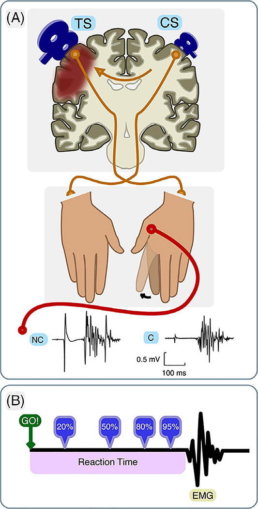
Objective: Patients with chronic stroke have been shown to have failure to release interhemispheric inhibition (IHI) from the intact to the damaged hemisphere before movement execution (premovement IHI). This inhibitory imbalance was found to correlate with poor motor performance in the chronic stage after stroke and has since become a target for therapeutic interventions. The logic of this approach, however, implies that abnormal premovement IHI is causal to poor behavioral outcome and should therefore be present early after stroke when motor impairment is at its worst. To test this idea, in a longitudinal study, we investigated interhemispheric interactions by tracking patients' premovement IHI for one year following stroke.

Methods: We assessed premovement IHI and motor behavior five times over a 1-year period after ischemic stroke in 22 patients and 11 healthy participants.

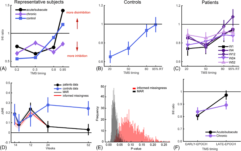
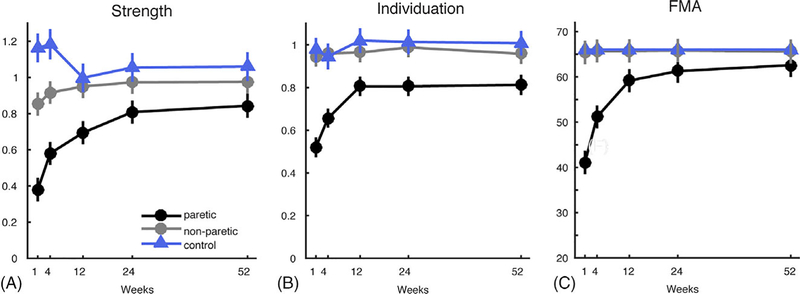
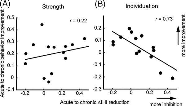
Results: We found that premovement IHI was normal during the acute/subacute period and only became abnormal at the chronic stage; specifically, release of IHI in movement preparation worsened as motor behavior improved. In addition, premovement IHI did not correlate with behavioral measures cross-sectionally, whereas the longitudinal emergence of abnormal premovement IHI from the acute to the chronic stage was inversely correlated with recovery of finger individuation.

Interpretation: These results suggest that interhemispheric imbalance is not a cause of poor motor recovery, but instead might be the consequence of underlying recovery processes. These findings call into question the rehabilitation strategy of attempting to rebalance interhemispheric interactions in order to improve motor recovery after stroke. Ann Neurol 2019;85:502-513.


Conflict of interest statement

Potential Conflicts of Interest

Nothing to report.

Figures

FIGURE 1:
FIGURE 1:
Schematic illustration of the premovement Interhemispheric Inhibition (IHI) paradigm. (A) A test stimulus (TS) was delivered over the lesioned hemisphere, and a conditioning stimulus (CS) was applied over the intact hemisphere before index finger abduction of the paretic hand (or right hand in healthy age-matched controls). In nonconditioned (NC) trials, only the TS was delivered, whereas in conditioned (C) trials, the CS preceded TS by 10 ms. EMG signals were recorded from the first dorsal interosseous muscle (FDI) of the moving hand. (B) TMS pulses were delivered at four timing epochs relative to the individual’s mean reaction time, estimated from a simple-reaction task. EMG = electromyography.
FIGURE 2:
FIGURE 2:
Lesion distribution of patients (N = 21). Averaged lesion distribution mapped to JHU-MNI space, with lesion flipped to one hemisphere. Color bar indicates patient count.
FIGURE 3:
FIGURE 3:
Release of IHI before movement onset. (A) IHI curves for a representative patient and a healthy control. These exemplar IHI profiles illustrate the normal release of IHI in patients at the acute/subacute stage, comparable to control subjects, and the lack of normal release of IHI during the chronic period. (B) Overall mean IHI curves for healthy controls. Because there were no differences over time in premovement IHI in controls (mixed-effects model with Week and TMS-Timing as fixed factors showed no significant effect of Week, χ2 = 0.067, p = 0.80, but significant main effect of TMS-Timing, χ2 = 22.28, p < 0.001), we averaged control data across weeks. (C) IHI curves for each time point over the 1-year period for patients. (D) Evolution of ΔIHI for patients and controls over the 1-year period. Patients showed close to control level of ΔIHI in the acute/subacute periods (W1–12), but their ΔIHIs became abnormal at the chronic stage. Shaded plots in gray and red are sensitivity analysis with two imputation schemes with MAR and informed-missingness cases, respectively, where missing not at random (MNAR) cases are imputed with 1,000 samples from N~(μ(t,patient), σ(t,patient)) or N~(0, σpatient). (μ(t,patient) and σ(t,patient)) are estimated from patients data at each time point and σpatient is estimated from all patients’ data. (E) Distribution of p values from sensitivity analysis with multiple imputation for the MAR and informed-missingness cases. (F) Change of IHI level at different movement preparation epochs in patients from the acute/subacute to chronic stage after stroke. There was a significant interaction of IHIEARLY-EPOCH vs IHILATE-EPOCH or acute/subacute and chronic stages (χ2 = 4.34, p = 0.037), but no differences when comparing across acute/subacute vs chronic stages for IHIEARLY-EPOCH (t(14) = 0.75; p = 0.47) or IHILATE-EPOCH (t(14) = 1.69; p = 0.11). Means and variances in all plots were estimated by mixed models. IHI = interhemispheric inhibition; TMS = transcranial magnetic stimulation.
FIGURE 4:
FIGURE 4:
Recovery curves for behavior measures of hand function over 1-year period, from week 1 to 52. (A) Strength indices. (B) Individuation indices. (C) FMA. Means and variances are estimated by mixed model. FMA = Fugl-Meyer Assessment.
FIGURE 5:
FIGURE 5:
Correlations between the reduction of premovement IHI (ΔIHI) from acute/subacute to chronic stages and the amount of behavioral recovery: (A) Strength; (B) Individuation. x- and y-axes are the mean differences between chronic and acute/subacute behavior measures and ΔIHI, respectively.
FIGURE 6:
FIGURE 6:
Other behavioral and physiological measures in premovement experiments. Reaction time (RT) for patients (A) and controls (B) at different TMS timing during movement preparation across the 1-year period. RTs for controls were overall faster than patients. Background EMG for patients (C) was overall lower than that in controls (D), but was at a similar level for conditioned vs nonconditioned TMS stimulation. RMS = root mean square; TMS = transcranial magnetic stimulation.

References

    1. Ward NS, Cohen LG. Mechanisms underlying recovery of motor function after stroke. Arch Neurol 2004;61:1844–1848. - PMC - PubMed
    1. Murase N, Duque J, Mazzocchio R, Cohen LG. Influence of interhemispheric interactions on motor function in chronic stroke. Ann Neurol 2004;55:400–409. - PubMed
    1. Nair DG, Renga V, Lindenberg R, et al. Optimizing recovery potential through simultaneous occupational therapy and non-invasive brain-stimulation using tDCS. Restor Neurol Neurosci 2011;29:411–420. - PMC - PubMed
    1. Lindenberg R, Renga V, Zhu LL, et al. Bihemispheric brain stimulation facilitates motor recovery in chronic stroke patients. Neurology 2010; 75:2176–2184. - PMC - PubMed
    1. Hummel FC, Cohen LG. Non-invasive brain stimulation: a new strategy to improve neurorehabilitation after stroke? Lancet Neurol 2006; 5:708–712. - PubMed

Publication types

MeSH terms