Review
. 2019 Mar;41(3):e1800198.
doi: 10.1002/bies.201800198.

Veins and Arteries Build Hierarchical Branching Patterns Differently: Bottom-Up versus Top-Down

Affiliations
Review

Veins and Arteries Build Hierarchical Branching Patterns Differently: Bottom-Up versus Top-Down

Kristy Red-Horse et al. Bioessays. 2019 Mar.

Abstract

A tree-like hierarchical branching structure is present in many biological systems, such as the kidney, lung, mammary gland, and blood vessels. Most of these organs form through branching morphogenesis, where outward growth results in smaller and smaller branches. However, the blood vasculature is unique in that it exists as two trees (arterial and venous) connected at their tips. Obtaining this organization might therefore require unique developmental mechanisms. As reviewed here, recent data indicate that arterial trees often form in reverse order. Accordingly, initial arterial endothelial cell differentiation occurs outside of arterial vessels. These pre-artery cells then build trees by following a migratory path from smaller into larger arteries, a process guided by the forces imparted by blood flow. Thus, in comparison to other branched organs, arteries can obtain their structure through inward growth and coalescence. Here, new information on the underlying mechanisms is discussed, and how defects can lead to pathologies, such as hypoplastic arteries and arteriovenous malformations.

Keywords: angiogenesis; arteries; blood flow; branching morphogenesis; coronary vasculature; cxcr4; notch; vein.


Figures

Figure 1.
Figure 1.
The vasculature consists of two interconnected trees. Schematized drawing of arterial and venous blood vessel trees. Note that the two trees are interconnected at their tips, allowing blood to flow from the arteries to the veins.
Figure 2.
Figure 2.
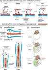
Sprouting angiogenesis at different developmental stages. A) Intersegmental blood vessel (ISV) sprouts emerge from the dorsal aorta in response to VEGF signaling, migrate dorsally and anastomose to form a network of arterial vessels that lacks a venous connection at this time point. Sprouting from the posterior cardinal vein in response to BMP signaling generates secondary sprouts (1.5 dpf). About half of the secondary sprouts connect to arterial ISVs at 2 dpf, allowing blood to flow from arteries into the venous circulation. Endothelial cells within arterial ISVs can change their identity to a venous fate or be replaced by venous cells through migration against the direction ofblood flow. dpf:days post fertilization. B) Angiogenesis in the zebrafish brain. New sprouts emerge from two laterally located veins (PHBC) and connect to a pre-existing artery (BA) located medially. Note expression of the chemokine Cxcl12b around the artery (orange). Sprouting tip cells sense Cxcl12b via the chemokine receptor cxcr4a and migrate toward the artery. PHBC: primordial hindbrain channel; BA: basilar artery. C: Live imaging of artery formation during fin regeneration in zebrafish. Vein-derived cells at the distal tip of the advancing vascular front change their direction of migration and become incorporated into newly forming arteries. This is dependent on the chemokine receptor Cxcr4a. Cxcl12 is also expressed in the vicinity of the artery. D) Genetic lineage labeling of sinus venosus (sv) endothelial cells at embryonic day (E) 10 (green) traces into the coronary vessel plexus and coronary arteries at later stages. Ao, aorta; pt, pulmonary trunk.
Figure 3.
Figure 3.
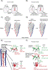
Comparison between current and newly proposed models for artery formation during blood vessel plexus remodeling. A) Current model of coronary plexus remodeling involves artery differentiation triggered by blood flow. B) Proposed model shows artery differentiation is initiated prior to the onset of blood flow via genetic pathways. This pre-specification of arterial fates would subsequently be refined via the ensuing blood flow. C) Current model of artery differentiation within the mouse retinal blood vessel plexus. Artery differentiation would occur in capillaries close to the existing artery, thereby expanding the artery. D) Proposed model involving pre-specification of arterial cells at the tip of the advancing vascular front. These would then grow toward the pre-existing artery, against the blood flow direction. E) Current model of the function of Notch signaling in the angiogenic front. VEGF leads to expression of the Notch ligand dll4 in tip cells. In turn, dll4 induces Notch signaling in neighboring stalk cells, preventing them from becoming a tip cell. Ensuing induction of Notch signaling leads to artery formation in locations deeper within the plexus. F) Proposed model linking the role of Notch signaling in angiogenesis and during artery formation. In this model, the tip cell initially displays low Notch signaling and expresses dll4. However, this does not lead to an induction of Notch signaling in the stalk cell for an unknown reason. Subsequently, some tip cells activate Notch signaling, presumably due to dll4 signaling from stalk cells. This leads to the onset of an arterial differentiation program that destines these tip cells to form new arteries. Tip cells change their direction of migration, possibly due to expression of the chemokine receptor cxcr4, and stop proliferating (possibly regulated via the transcription factor COUP-TFIII). Finally, cells before present at the tip position connect to a pre-existing artery. In this model, initiation of artery formation occurs in tip cells at the angiogenic front. Both models are not necessarily mutually exclusive.
Figure 4.
Figure 4.
Function of genes involved in cell migration during artery formation. A) In vitro data showing that overexpression of Dach1 leads to an increased response to shear stress, causing enhanced endothelial cell migration against the direction of the flowing media. Cells lacking Endoglin or SMAD4 function show an impaired response to the flowing media and fail to migrate properly. B) Cdh5-CreERT2-mediated activation of GFP lineage marker within endothelial cells of the mouse retina reveals an equal contribution to arteries, veins, and capillaries. Deletion of the BMP co-receptor Endoglin results in fewer cells in tip position and in arteries, while overexpression of Endoglin causes cells to contribute more to arteries, an effect that is amplified by a deletion of Endoglin in the surrounding retinal endothelial cells. Deletion of the small GTPase cdc42 leads to an accumulation of mutant cells within and around veins, while mutant cells are less abundant at the angiogenic front and in arteries. C) Summary of cellular behaviors within arterial and venous blood vessel trees that might contribute to their hierarchical patterning. In both trees, endothelial cells migrate against the direction of the flowing blood (indicated by arrows).This distributes cells from smaller, more distally located segments toward larger proximal vessels. Endothelial cells within arteries show low proliferation; thus redistribution of cells necessary for assigning proper blood vessel calibers can only occur via migration. Endothelial cells within venous blood vessel trees proliferate and migrate distally toward smaller branches. Here, some cells become genetically specified as pre-artery cells, which subsequently build newly forming arteries.

Similar articles

Cited by

References

    1. Blake J, Rosenblum ND, Semin. Cell Dev. Biol. 2014, 36, 2. - PubMed
    1. Hayashi S, Kondo T, Genetics 2018, 209, 367. - PMC - PubMed
    1. Ochoa-Espinosa A, Affolter M, Cold Spring Harb. Perspect. Biol. 2012, 4, a008243. - PMC - PubMed
    1. Short KM, Smyth IM, Nat. Rev. Nephrol. 2016, 12, 754. - PubMed
    1. Varner VD, Nelson CM, Development 2014, 141, 2750. - PMC - PubMed

Publication types

Substances