. 2019 Jul 1;28(13):2120-2132.
doi: 10.1093/hmg/ddz044.

Sunitinib promotes myogenic regeneration and mitigates disease progression in the mdx mouse model of Duchenne muscular dystrophy

Affiliations

Sunitinib promotes myogenic regeneration and mitigates disease progression in the mdx mouse model of Duchenne muscular dystrophy

Tatiana M Fontelonga et al. Hum Mol Genet. .

Abstract

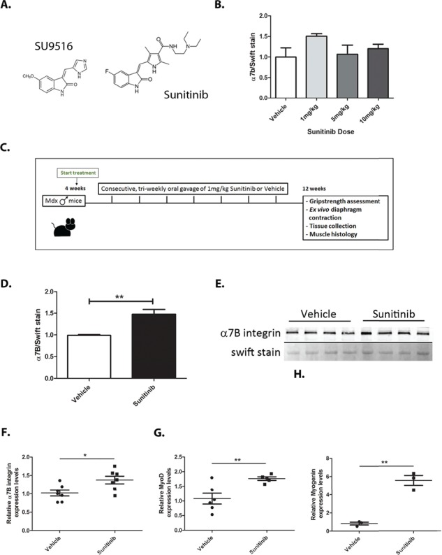
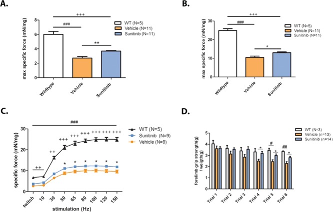
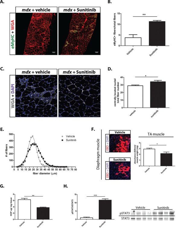
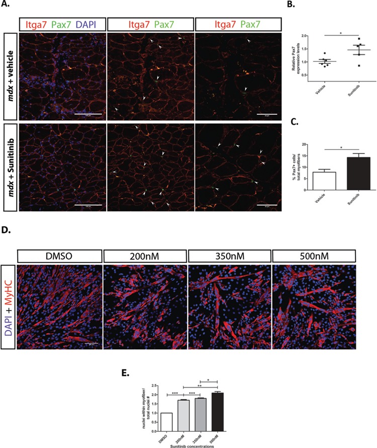
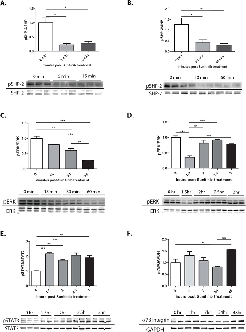
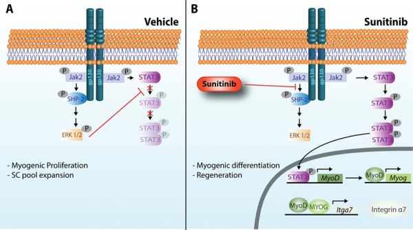
Duchenne muscular dystrophy (DMD) is a lethal, muscle degenerative disease causing premature death of affected children. DMD is characterized by mutations in the dystrophin gene that result in a loss of the dystrophin protein. Loss of dystrophin causes an associated reduction in proteins of the dystrophin glycoprotein complex, leading to contraction-induced sarcolemmal weakening, muscle tearing, fibrotic infiltration and rounds of degeneration and failed regeneration affecting satellite cell populations. The α7β1 integrin has been implicated in increasing myogenic capacity of satellite cells, therefore restoring muscle viability, increasing muscle force and preserving muscle function in dystrophic mouse models. In this study, we show that a Food and Drug Administration (FDA)-approved small molecule, Sunitinib, is a potent α7 integrin enhancer capable of promoting myogenic regeneration by stimulating satellite cell activation and increasing myofiber fusion. Sunitinib exerts its regenerative effects via transient inhibition of SHP-2 and subsequent activation of the STAT3 pathway. Treatment of mdx mice with Sunitinib demonstrated decreased membrane leakiness and damage owing to myofiber regeneration and enhanced support at the extracellular matrix. The decreased myofiber damage translated into a significant increase in muscle force production. This study identifies an already FDA-approved compound, Sunitinib, as a possible DMD therapeutic with the potential to treat other muscular dystrophies in which there is defective muscle repair.


Figures

Figure 1
Figure 1
Sunitinib treatment increases α1;7B integrin via activation of Myod1 and Myog transcription factors. (A) Structural similarities between the known integrin α7 enhancing molecule SU9516 and Sunitinib. (B) Western blot dose response curve performed on N = 4 mdx mice with daily 1 mg/kg–10 mg/kg Sunitinib treatment for a total of 5 days showing optimal α7B integrin enhancing dose at 1 mg/kg. (C) Schematic representation of the 8 week Sunitinib treatment plan and final muscle assessments. (D and E) Western blot analysis and quantification of diaphragm α7B integrin levels showing a 1.5-fold increase in α7B expression with Sunitinib treatment. (F) RT-qPCR analysis of α7B integrin transcript levels showing ~1.4-fold increase with Sunitinib treatment. (G and H) RT-qPCR analysis of α7B upstream transcription factors and differentiation markers MyoD1 and Myog showing a ~1.8-fold and ~5.8-fold transcript increase, respectively, with Sunitinib treatment. Statistical significance of mean ± SEM; *P < 0.05, **P < 0.01.
Figure 2
Figure 2
Sunitinib improves specific muscle contractile function and overall muscle strength. Muscle isometric contractility and strength assessments performed on 12-week-old mdx mice. (A) Single 1 Hz pulses generated isometric twitch force outputs showing the severe decline in diaphragm muscle twitch between WT (N = 5) and mdx vehicle-treated muscle (N = 11); Sunitinib-treated muscle (N = 11) twitch is significantly increased compared to vehicle treated. (B) Isometric tetanic stimuli performed at 100 Hz on diaphragm muscle showing a significant decline in tetanic force output between WT (N = 5) and vehicle-treated muscle (N = 11), Sunitinib treatment (N = 11) increased isometric tetanic force compared to vehicle treatment. (C) Isometric force performed at increasing stimulation frequencies; Sunitinib treatment increased force production in the 50–150 Hz frequency stimulations when compared to vehicle-treated muscle. (D) Muscle exhaustion depicted as decreased forelimb grip strength is apparent in vehicle-treated mdx mice compared to WT mice at trials 5 and 6. No significant change in forelimb force is observed between WT and Sunitinib-treated mdx mice during the six trials; Sunitinib-treated mdx mice are stronger than the vehicle treated in trials 4–6 suggesting less muscle exhaustion. Data assessed for significance using one-way ANOVA and statistical significance of mean ± SEM; WT versus mdx vehicle treatment #P < 0.05, ##P < 0.01, ###P < 0.001; WT versus mdx-Sunitinib treatment ++P < 0.01, +++P < 0.001; mdx vehicle treatment versus mdx-Sunitinib treatment *P < 0.05, **P < 0.01.
Figure 3
Figure 3
Sunitinib promotes muscle repair and improves markers of DMD disease progression. (A) Representative section of immunohistochemistry performed on 10 μm diaphragm muscle sections showing increased number of eMyHC (green) positive fibers in Sunitinib-treated muscle. WGA (red); scale bar, 100 μm. (B) Quantification of eMyHC positive (+) muscle fibers normalized to total fiber numbers of a whole mdx diaphragm muscle section (N = 4); Sunitinib treatment causes ~8% increase in eMyHC+ fibers compared to vehicle treatment. (C and D) CLN counts performed on WGA (grey) and DAPI (blue) immunolabeled 10 μm diaphragm muscle sections (N = 6); Sunitinib treatment increased CLNs by ~5% compared to vehicle treated. (E) Myofiber size distribution of Sunitinib- and vehicle-treated whole 10 μm sections diaphragm sections, measured using minimum Feret’s diameter (N = 4); Sunitinib-treated muscle shows a shift toward higher percentage of large fiber sizes compared to vehicle treated. (F) Representative sections of EBD (red) infiltration in Sunitinib-treated diaphragm muscle. EBD quantification performed on total GA muscle shows decreased EBD infiltration compared to vehicle treated (N = 4). (G) Hydroxyproline assay was performed to quantify the amount of fibrotic infiltration in whole GA muscle (N = 4); Sunitinib treatment decreases fibrosis as shown by a 2-fold decrease in collagen content in the mdx GA muscle, compared to vehicle treated. (H) Western blot analysis showing the STAT3 pathway is activated in response to Sunitinib treatment 1 h post final dose (6 week total dosing) in mdx diaphragm muscle. Data assessed for significance using unpaired t-test and statistical significance of mean ± SEM; *P < 0.05, **P < 0.01, ***P < 0.001.
Figure 4
Figure 4
Sunitinib treatment promotes SC proliferation and myoblast fusion. (A) Immunohistochemistry performed on diaphragm muscle sections of mdx mice treated with vehicle or Sunitinib. Higher numbers of SCs are present in Sunitinib-treated diaphragm; SCs are identified (arrows) by their location around myofibers [α7B integrin (red)], Pax7+ cells (green) and co-localization with nuclear DAPI (blue) stain. Middle panels without DAPI staining for easier identification of SCs. Panels to the right were taken at 60× magnification, also to better identify SCs. (B) Increased Pax7 transcript levels observed in Sunitinib-treated TA whole muscle. (C) Quantification of SC numbers, determined by counting Pax7+ cells per total fiber counts (10 panels per tissue, magnification 40×, N = 3); Sunitinib treatment significantly increased SC numbers in diaphragm and TA muscles. (D) C2C12 myoblasts under differentiating conditions 48 h post DMSO or Sunitinib treatment at varying concentrations immunolabeled for MHC (red) and nuclear DAPI (blue). (E) Fusion index determined by counting the number of nuclei within MHC+ fibers and normalizing to total nuclei counts per 10× panel (N = 3); Increased myofiber fusion observed at all three Sunitinib concentrations with 500 nm showing the highest fusion index compared to DMSO-vehicle-treated cells. Data assessed for significance using one-way ANOVA and statistical significance of mean ± SEM; *P < 0.05, **P < 0.01, ***P < 0.001
Figure 5
Figure 5
Sunitinib inhibits SHP-2–ERK1/2 and activates the STAT3 pathway in C2C12 cell line. C2C12 myoblasts treated with 500 nm Sunitinib in triplicate (N = 3) and taken at different time points to assess SHP-2–ERK1/2 inhibition and STAT3 activation. (A) Quantification of SHP-2 phosphorylation shows significant inhibition at 5 min lasting up to 15 min. (B) Quantification of SHP-2 phosphorylation shows significant inhibition continuing from 30 min to 1 h. (C) Quantification of ERK1/2 phosphorylation shows significant inhibition starting at 15 min post-treatment and lasting up to 1 h. (D) Quantification of ERK1/2 phosphorylation showing continued inhibition at 1.5 h with recuperating phosphorylation after 2 h of treatment. (E) Quantification of STAT3 phosphorylation showing activation after 1.5 h of treatment. (F) Quantification of α7B integrin shows a 1.5-fold increase in expression 48 h post-treatment. Data assessed for significance using one-way ANOVA and statistical significance of mean ± SEM; *P < 0.05, **P < 0.01, ***P < 0.001.
Figure 6
Figure 6
Proposed mechanism of SHP2–ERK1/2 inhibition and STAT3 activation by Sunitinib. In response to muscle injury, gp130 mediated by Jak2 can activate both ERK1/2 and STAT3. (A) Upon gp130 receptor dimerization in response to growth factor binding, constitutively bound Jak2 is activated and trans-phosphorylates gp130 at several tyrosine residues (Y). P-Y759 recruits SHP-2 that is phosphorylated by Jak2 and signals via Grb2-Sos-Ras–Raf and MEK to activate ERK1/2. Once activated, ERK1/2 has been shown to directly phosphorylate STAT3 at a serine residue, thus preventing phosphorylation at tyrosine residues and nuclear translocation (50). ERK1/2 becomes the predominant signaling pathway promoting the proliferation of myogenic cells. (B) Sunitinib inhibits the activation of ERK1/2, potentially by (1) preventing the association of SHP-2 with gp130 and (2) preventing the phosphorylation of SHP-2 by Jak2. Non-phosphorylated ERK1/2 no longer phosphorylates STAT3 at its serine residue, thus allowing tyrosine phosphorylation and activation of STAT3 by Jak2. Dimerized STAT3 translocates into the nucleus where it promotes the transcription of the transcription factor MyoD1 that can subsequentially promote the transcription of myogenin (Myog). Both MyoD1 and MYOG can then promote the transcription of integrin α7 (Itga7). Integrin α7 promotes differentiation and fusion of myofibers, enhancing muscle regeneration.

Similar articles

Cited by

References

    1. Yiu E.M. and Kornberg A.J. (2015) Duchenne muscular dystrophy. J. Paediatr. Child Health, 51, 759–764. - PubMed
    1. Hoffman E.P., Brown R.H. and Kunkel L.M. (1987) Dystrophin: the protein product of the Duchenne muscular dystrophy locus. Cell, 51, 919–928. - PubMed
    1. Bonilla E., Samitt C.E., Miranda A.F., Hays A.P., Salviati G., DiMauro S., Kunkel L.M., Hoffman E.P. and Rowland L.P. (1988) Duchenne muscular dystrophy: deficiency of dystrophin at the muscle cell surface. Cell, 54, 447–452. - PubMed
    1. Durbeej M. and Campbell K.P. (2002) Muscular dystrophies involving the dystrophin–glycoprotein complex: an overview of current mouse models. Curr. Opin. Genet. Dev., 12, 349–361. - PubMed
    1. Le Rumeur E., Winder S.J. and Hubert J.F. (2010) Dystrophin: more than just the sum of its parts. Biochim. Biophys. Acta Proteins Proteom., 1804, 1713–1722. - PubMed

Publication types

MeSH terms