. 2019 Apr 15:486:79-88.
doi: 10.1016/j.mce.2019.02.018. Epub 2019 Feb 23.

Adrenal androgens rescue prostatic dihydrotestosterone production and growth of prostate cancer cells after castration

Affiliations

Adrenal androgens rescue prostatic dihydrotestosterone production and growth of prostate cancer cells after castration

Yue Wu et al. Mol Cell Endocrinol. .

Abstract

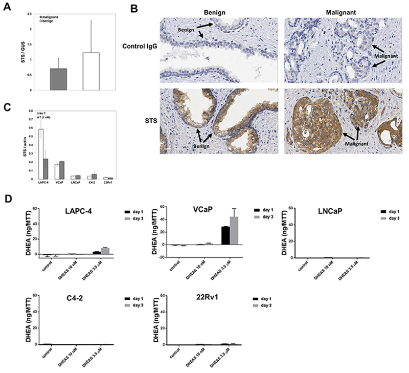
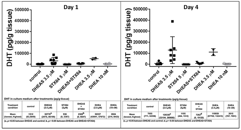
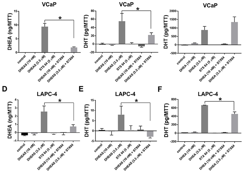
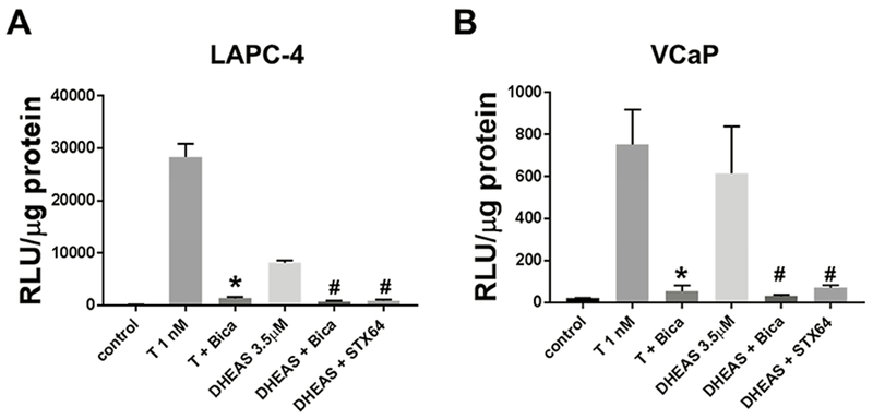
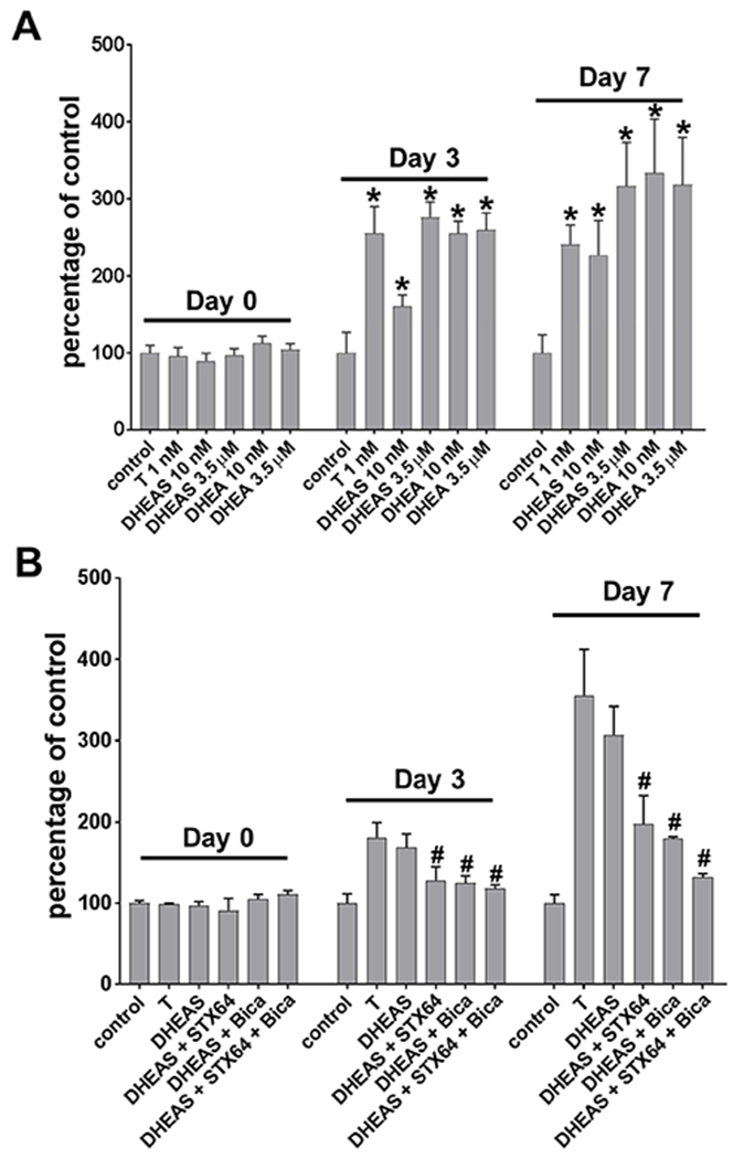
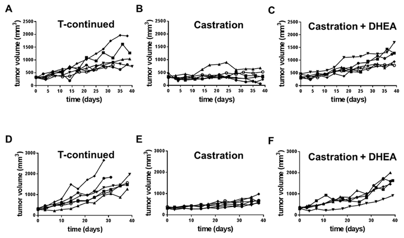
Adrenal androgens dehydroepiandrosterone (DHEA) and DHEA-sulfate (DHEAS) are potential substrates for intracrine production of testosterone (T) and dihydrotestosterone (DHT), or directly to DHT, by prostate cancer (PCa) cells. Production of DHT from DHEAS and DHEA, and the role of steroid sulfatase (STS), were evaluated ex vivo using fresh human prostate tissue and in vitro using human PCa cell lines. STS was expressed in benign prostate tissue and PCa tissue. DHEAS at a physiological concentration was converted to DHT in prostate tissue and PCa cell lines, which was STS-dependent. DHEAS activation of androgen receptor (AR) and stimulation of PCa cell growth were STS-dependent. DHEA at a physiological concentration was not converted to DHT ex vivo and in vitro, but stimulated in vivo tumor growth of the human PCa cell line, VCaP, in castrated mice. The findings suggest that targeting metabolism of DHEAS and DHEA may enhance androgen deprivation therapy.

Keywords: Adrenal androgen; Androgen; Intracrine androgen metabolism; Metabolism; Prostate cancer; Steroid sulfatase.


Conflict of interest statement

Conflict of interest: The authors declare no potential conflicts of interest.

Figures

Fig 1.
Fig 1.
Expression of STS in human prostate cancer tissue and human prostate cancer cell lines, and the conversion of DHEAS to DHEA by prostate cancer cell lines (A) STS expression in benign human prostate tissue and prostate cancer tissue at mRNA levels (n=20); (B) STS expression at protein levels in matched benign and malignant prostate tissue specimens; (C) STS expression at mRNA levels in human prostate cancer cell lines; (D) DHEA production by prostate cancer cell lines treated with DHEAS for 1 day or 3 days. DHEA in the culture medium was normalized against cell numbers, which was indicated by units of OD570 measured using the MTT assay.
Fig 2.
Fig 2.
DHT production by prostate tissue ex vivo was STS-dependent. Inserted tables showed the exact values of DHT production, in each treatment presented in the respective bar graph at the top in the format of mean and range (lowest value, highest value). DHEAS 3.5 μM and STX64 5 μM were used in the combination treatments.
Fig 3.
Fig 3.
DHEA and DHT production by prostate cancer cell lines VCaP (Panels A-C) and LAPC-4 (Panels D-F) using DHEAS was STS-dependent. (A) & (D), DHEA production; (B) & (E), DHT production by cells treated with DHEAS; (C) & (F), DHT production by cells treated with DHEAS. Cells were treated for 3 days. Androgens in the culture medium were normalized against cell numbers, which was indicated by units of OD570 measured using the MTT assay. * p < 0.05.
Fig 4.
Fig 4.
DHEAS activation of AR in LAPC-4 and VCaP cell lines was diminished by AR antagonist bicalutamide (Bica) and STX64. T 1 nM, Bicalutamide 10 μM, DHEAS 3.5 μM, and STX64 5 μM were used in the combination treatments. Cells were treated for 24 hr. * p < 0.05 compared to T 1 nM. # p < 0.05 compared to DHEAS 3.5 μM.
Fig 5.
Fig 5.
AR-dependent stimulation of growth by adrenal androgens. (A) DHEAS and DHEA stimulated growth of VCaP cells. (B) DHEAS (DS) stimulated growth was diminished by AR antagonist bicalutamide (Bica) and STX64 (B). Cells were treated for 3 days and 7 days. Growth was assessed using MTT assay. Data were presented in percentage to untreated controls at each time point (day). * p < 0.05 compared to control. # p < 0.05 compared to DHEAS.
Fig 6.
Fig 6.
DHEA sustained growth of VCaP xenograft after castration in SCID mice (A-C) and nude mice (E-F). Each line represented the growth curve of the xenograft in one mouse.

Similar articles

Cited by

References

    1. Dai C, Heemers H and Sharifi N, 2017. Androgen signaling in prostate cancer, Cold Spring Harb Perspect Med. 7 10.1101/cshperspect.a030452 - DOI - PMC - PubMed
    1. Stuchbery R, McCoy PJ, Hovens CM and Corcoran NM, 2017. Androgen synthesis in prostate cancer: do all roads lead to Rome?, Nat Rev Urol 14, 49–58. 10.1038/nrurol.2016.221 - DOI - PubMed
    1. Mohler JL, 2008. A role for the androgen-receptor in clinically localized and advanced prostate cancer, Best Pract Res Clin Endocrinol Metab 22, 357–72. 10.1016/j.beem.2008.01.009 - DOI - PMC - PubMed
    1. Wilson EM and French FS, 1976. Binding properties of androgen receptors. Evidence for identical receptors in rat testis, epididymis, and prostate, J Biol Chem 251, 5620–9. - PubMed
    1. Kamrath C, Hochberg Z, Hartmann MF, Remer T and Wudy SA, 2012a. Increased activation of the alternative “backdoor” pathway in patients with 21-hydroxylase deficiency: evidence from urinary steroid hormone analysis, J Clin Endocrinol Metab 97, E367–75. 10.1210/jc.2011-1997 - DOI - PubMed

Publication types