Review
. 2019 Feb:54:86-94.
doi: 10.1016/j.sbi.2019.01.003. Epub 2019 Feb 23.

Regulatory mechanisms in postsynaptic phosphorylation networks

Affiliations
Review

Regulatory mechanisms in postsynaptic phosphorylation networks

Marcelo P Coba. Curr Opin Struct Biol. 2019 Feb.

Abstract

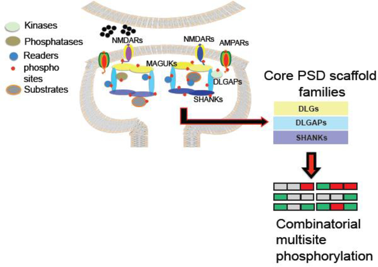
The modulation of the postsynaptic signaling machinery by protein phosphorylation has attracted much interest since it is key for the understanding of the regulation of a variety of synaptic functions. While advances in mass spectrometry have allowed us to begin performing large-scale analysis of protein phosphorylation in components of the PSD, the systematic collection of datasets and their functional significance within the context of regulatory signaling networks is in its infancy. Here, we will focus on the composition of the PSD phosphoproteome describing kinase, phosphatase, and protein domain modules involved in the regulation of phosphorylation signaling. We will discuss the impact of synaptic plasticity mechanisms such as long-term potentiation (LTP) in mammalian kinomes and describe the general rules of signaling organization in the PSD phosphoproteome.


Figures

Figure 1
Figure 1
Schematic representation of PSD phosphorylation, showing kinase, phosphatase and phosphorylation readers. Cartoon shows core components of the PSD scaffold machinery. Top layer Disk Large associated (DLG) family members, also known as MAGUKs (see main text), middle scaffold layer composed of Disk Large Associated Guanylate Associated Proteins (DLGAPs) and a bottom layer of SHANK family scaffolds [,–13].
Figure 2
Figure 2
A (top). Representation of PSD protein kinases present in the mouse kinome. Circles show kinases present in different families of the kinome tree. Main groups are indicated: AGC, CMCG, TK, STE, and CAMK. Color code for each kinase represent if kinases shows increase, decrease or not known changes in protein phosphorylation and activity after the induction of LTP. Bottom table shows kinase families in the mouse kinome, number of kinases presents in the mouse PSD for each family, number of phosphorylation sites reported for each kinase family and number of phosphorylation sites that increase or decrease after the induction of LTP. B (top). Figure shows the composition of the mouse phosphatome and mapping of PSD phosphatases within the mouse phosphatome tree. Bottom table shows protein domains present in PSD proteins that have the capacity to recognize and bind to phosphorylated protein sequences (readers). Table shows domain ID (SMART database), domain name, number of PSD proteins containing each domain, if the domain is enriched in PSD fractions and phosphorylation specificity of each reader module.
Figure 3
Figure 3
Regulatory motifs in multiple-phosphorylated sequences, with one kinase (k) phosphorylating multiple sites (a), different kinases converging in multiple phosphorylated regions (b). These kinase interaction can prime (enhance) the phosphorylation of nearby sites (c) or impair the phosphorylation by a second kinase (d). Figure 3e and f shows convergence of AGC kinases such as PKC, PKA, PKG, AKT with basophilic kinases from CAMK family like CAMK2, TRIO, DAPK. Overlapping multiphosphorylation sequences in glutamate receptors driven by activation of NMDA, mGlu and dopamine receptors (top 3 panels). Cartoon shows changes in phosphorylation observed in different subunits of NMDAR (GRIN1, GRIN2B) and AMPA (GRIA1) receptors. Lower 3 panels shows kinases involved in the phosphorylation of each site/sequence.

References

    1. Bayes A, van de Lagemaat LN, Collins MO, Croning MD, Whittle IR, Choudhary JS, Grant SG: Characterization of the proteome, diseases and evolution of the human postsynaptic density. Nat Neurosci 2011, 14:19–21. - PMC - PubMed
    1. Collins MO, Husi H, Yu L, Brandon JM, Anderson CN, Blackstock WP, Choudhary JS, Grant SG: Molecular characterization and comparison of the components and multiprotein complexes in the postsynaptic proteome. J Neurochem 2006, 97 Suppl 1:16–23. - PubMed
    1. Cheng D, Hoogenraad CC, Rush J, Ramm E, Schlager MA, Duong DM, Xu P, Wijayawardana SR, Hanfelt J, Nakagawa T, et al. : Relative and absolute quantification of postsynaptic density proteome isolated from rat forebrain and cerebellum. Mol Cell Proteomics 2006, 5:1158–1170. - PubMed
    1. Chen X, Vinade L, Leapman RD, Petersen JD, Nakagawa T, Phillips TM, Sheng M, Reese TS: Mass of the postsynaptic density and enumeration of three key molecules. Proc Natl Acad Sci U S A 2005, 102:11551–11556. - PMC - PubMed
    1. Sheng M, Kim E: The postsynaptic organization of synapses. Cold Spring Harb Perspect Biol 2011, 3. - PMC - PubMed

Publication types

Substances

LinkOut - more resources