

Changes in Mannose-Binding Lectin and Collectin Kidney 1 Levels in Sepsis Patients With and Without Disseminated Intravascular Coagulation
- PMID: 30808212
- PMCID: PMC6714923
- DOI: 10.1177/1076029618821189
Changes in Mannose-Binding Lectin and Collectin Kidney 1 Levels in Sepsis Patients With and Without Disseminated Intravascular Coagulation
Abstract
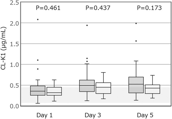
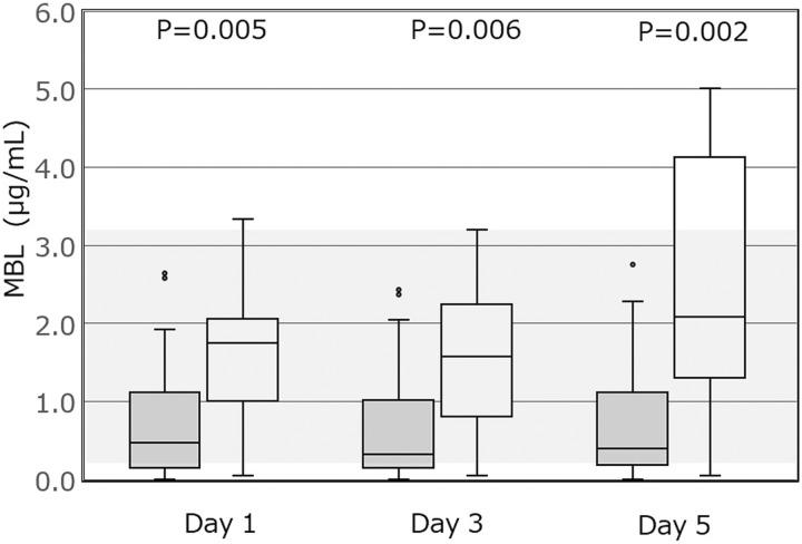
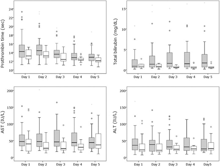
In sepsis, systemic coagulation activation frequently causes disseminated intravascular coagulation (DIC), and the uncontrolled activation of the complement system can induce multiple organ dysfunction and poor prognosis. This study aimed to examine the association of DIC with levels of collectin kidney 1 (CL-K1), a novel collectin of the complement system, and mannose-binding lectin (MBL), a classical-type collectin in patients with sepsis. We collected blood samples prospectively from adult patients with sepsis admitted to the intensive care unit (ICU) from day 1 (admission) to day 5. The CL-K1 and MBL levels were measured by enzyme-linked immunosorbent assay, and DIC was diagnosed by using a scoring algorithm. The correlation of CL-K1 and MBL levels with other coagulation markers was analyzed. There were 37 patients with DIC (DIC group) and 15 without DIC (non-DIC group). Compared to the non-DIC group, the DIC group had more severe conditions and higher mortality. During the 5 days after ICU admission, plasma CL-K1 levels were similar between the groups, but plasma MBL levels were significantly lower in the DIC group. Plasma CL-K1 levels were weakly correlated with prothrombin time, activated partial thromboplastin time, and antithrombin levels; plasma MBL levels were weakly correlated with fibrin/fibrinogen degradation product levels and DIC score. In conclusion, during the first 5 days of ICU admission, plasma CL-K1 levels were similar between the DIC and non-DIC groups. However, plasma MBL levels were lower in the DIC group compared to the non-DIC group, and the significance of this difference grew gradually over time.
Keywords: collectin; complement system proteins; disseminated intravascular coagulation; mannose-binding lectin; sepsis.
Conflict of interest statement
Figures
Similar articles
-
Elevated plasma CL-K1 level is associated with a risk of developing disseminated intravascular coagulation (DIC).J Thromb Thrombolysis. 2014 Oct;38(3):331-8. doi: 10.1007/s11239-013-1042-5. J Thromb Thrombolysis. 2014. PMID: 24474086 Free PMC article. Clinical Trial.
-
Predictive value of the complement system for sepsis-induced disseminated intravascular coagulation in septic patients in emergency department.J Crit Care. 2015 Apr;30(2):290-5. doi: 10.1016/j.jcrc.2014.11.007. Epub 2014 Nov 18. J Crit Care. 2015. PMID: 25547047
-
Reduced Mannose-Binding Lectin-Associated Serine Protease (MASP)-1 is Associated with Disturbed Coagulation in Septic Shock.Thromb Haemost. 2019 Jun;119(6):952-961. doi: 10.1055/s-0039-1685140. Epub 2019 Apr 15. Thromb Haemost. 2019. PMID: 30986866
-
A journey through the lectin pathway of complement-MBL and beyond.Immunol Rev. 2016 Nov;274(1):74-97. doi: 10.1111/imr.12468. Immunol Rev. 2016. PMID: 27782323 Review.
-
[A novel molecular marker for thrombus formation and life prognosis--clinical usefulness of measurement of soluble fibrin monomer-fibrinogen complex (SF)].Rinsho Byori. 2004 Apr;52(4):355-61. Rinsho Byori. 2004. PMID: 15164605 Review. Japanese.
Cited by
-
Early control of viral load by favipiravir promotes survival to Ebola virus challenge and prevents cytokine storm in non-human primates.PLoS Negl Trop Dis. 2021 Mar 29;15(3):e0009300. doi: 10.1371/journal.pntd.0009300. eCollection 2021 Mar. PLoS Negl Trop Dis. 2021. PMID: 33780452 Free PMC article.
References
-
- Angus DC, van der Poll T. Severe sepsis and septic shock. N Engl J Med. 2013;369(9):840–851. - PubMed
-
- Hunt BJ. Bleeding and coagulopathies in critical care. N Engl J Med. 2014;370(9):847–859. - PubMed
-
- Ogura H, Gando S, Saitoh D, et al. Epidemiology of severe sepsis in Japanese intensive care units: a prospective multicenter study. J Infect Chemother. 2014;20(3):157–162. - PubMed
-
- Keragala CB, Draxler DF, McQuilten ZK, Medcalf RL. Haemostasis and innate immunity – a complementary relationship: a review of the intricate relationship between coagulation and complement pathways. Br J Haematol. 2018;180(6):782–798. - PubMed
MeSH terms
Substances
LinkOut - more resources
Full Text Sources
Miscellaneous
