

miR-195-5p/NOTCH2-mediated EMT modulates IL-4 secretion in colorectal cancer to affect M2-like TAM polarization
- PMID: 30808369
- PMCID: PMC6390326
- DOI: 10.1186/s13045-019-0708-7
miR-195-5p/NOTCH2-mediated EMT modulates IL-4 secretion in colorectal cancer to affect M2-like TAM polarization
Erratum in
-
Correction to: miR-195-5p/NOTCH2-mediated EMT modulates IL-4 secretion in colorectal cancer to affect M2-like TAM polarization.J Hematol Oncol. 2019 Nov 22;12(1):122. doi: 10.1186/s13045-019-0810-x. J Hematol Oncol. 2019. PMID: 31757211 Free PMC article.
Retraction in
-
Retraction Note: miR-195-5p/NOTCH2-mediated EMT modulates IL-4 secretion in colorectal cancer to affect M2-like TAM polarization.J Hematol Oncol. 2023 Apr 19;16(1):41. doi: 10.1186/s13045-023-01438-0. J Hematol Oncol. 2023. PMID: 37076859 Free PMC article. No abstract available.
Abstract
Background: Tumor microenvironment (TME) is a complex environment containing tumor cells, tumor-associated macrophages (TAMs), interstitial cells, and non-cellular components. Epithelial-mesenchymal transition (EMT), as a major actor in cancer tumorigenicity and metastasis, was involved in the interaction between TAMs and tumor cells. However, the potential mechanisms of EMT and how EMT-programmed tumor cells affect M2-like TAMs still need further exploration.
Methods: An integrated analysis of nine CRC miRNA expression datasets was performed. Functional assays, including the EdU, clone formation, wound healing, and transwell assays, were used to determine the anticancer role of miR-195-5p in human CRC progression. Furthermore, RNA immunoprecipitation, RNA decay, and dual-luciferase reporter assays were used to determine the mechanism of miR-195-p CRC progression. Then co-culture, migration, and ELISA assays were applied to determine the role of miR-195-5p in macrophage recruitment and alternative polarization. Xenograft mouse models were used to determine the role of miR-195-5p in CRC tumorigenicity and TAM polarization in vivo.
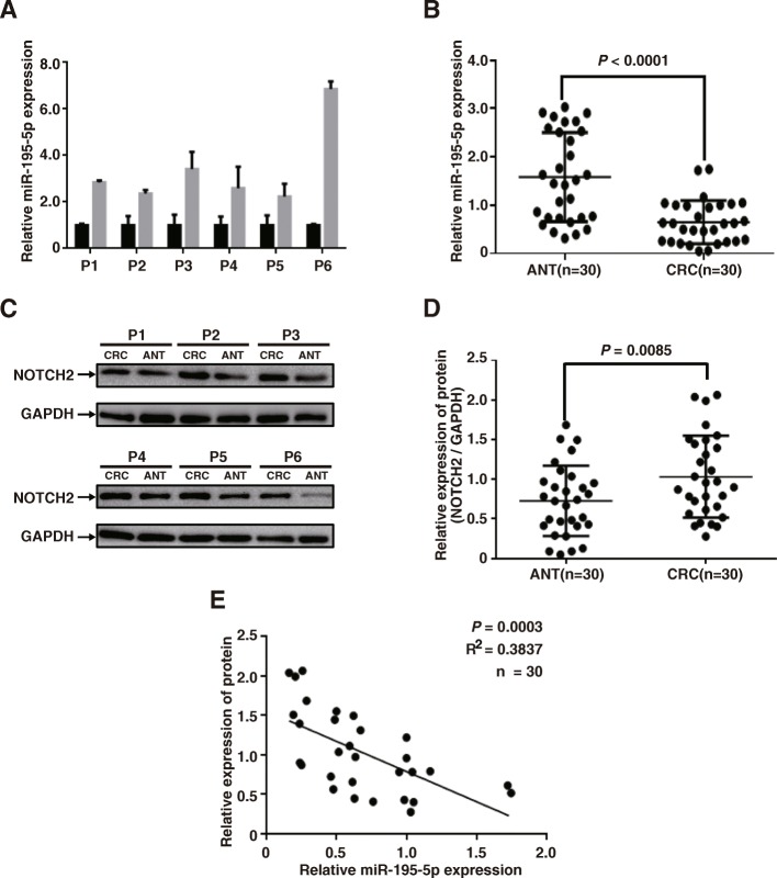
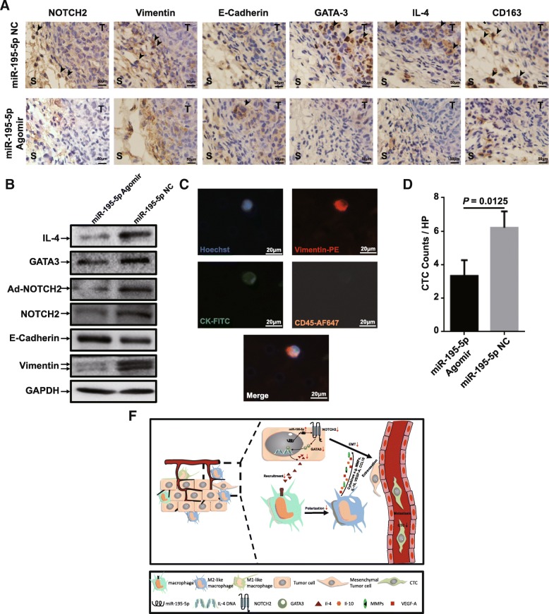
Results: An integrated analysis confirmed that miR-195-5p was significantly downregulated in CRC tissues, and patients with a low level of miR-195-5p had significantly shortened overall survival as revealed by the TCGA-COAD dataset. Altered miR-195-5p in colon cancer cells led to distinct changes of proliferation, migration, invasion, and EMT. Mechanistically, miR-195-5p regulated NOTCH2 expression in a post-transcriptional manner by directly binding to 3'-UTR of the Notch2 mRNA. Subsequently, miR-195-5p/NOTCH2 suppressed GATA3-mediated IL-4 secretion in CRC cells and ultimately inhibited M2-like TAM polarization.
Conclusions: miR-195-5p may play a vital role in regulating NOTCH2-mediated tumor cell EMT, thereby affecting IL-4-related M2-like TAM polarization in CRC.
Keywords: CRC; EMT; IL-4; NOTCH2; TAMs; miR-195-5p.
Conflict of interest statement
Ethics approval and consent to participate
The study was endorsed by the Research Ethics Committee of Wuhan University. Informed consents were obtained from all participating patients. This study complied with the Animal Care guidelines of Wuhan University.
Consent for publication
Informed consent for publication was obtained from all participants.
Competing interests
The authors declare that they have no competing interests.
Publisher’s Note
Springer Nature remains neutral with regard to jurisdictional claims in published maps and institutional affiliations.
Figures


Similar articles
-
MiR-135a-5p/STAT6-mediated EMT regulates IL-4 secretion in non-small cell lung cancer to affect M2-like TAM polarization.Int Immunopharmacol. 2025 May 16;155:114623. doi: 10.1016/j.intimp.2025.114623. Epub 2025 Apr 10. Int Immunopharmacol. 2025. PMID: 40199139
-
Tumor-derived exosomal microRNA-106b-5p activates EMT-cancer cell and M2-subtype TAM interaction to facilitate CRC metastasis.Mol Ther. 2021 Jun 2;29(6):2088-2107. doi: 10.1016/j.ymthe.2021.02.006. Epub 2021 Feb 9. Mol Ther. 2021. PMID: 33571679 Free PMC article.
-
Crosstalk between cancer cells and tumor associated macrophages is required for mesenchymal circulating tumor cell-mediated colorectal cancer metastasis.Mol Cancer. 2019 Mar 30;18(1):64. doi: 10.1186/s12943-019-0976-4. Mol Cancer. 2019. PMID: 30927925 Free PMC article.
-
A new perspective on the therapeutic potential of tumor metastasis: targeting the metabolic interactions between TAMs and tumor cells.Int J Biol Sci. 2024 Sep 23;20(13):5109-5126. doi: 10.7150/ijbs.99680. eCollection 2024. Int J Biol Sci. 2024. PMID: 39430253 Free PMC article. Review.
-
Regulation and Function of Tumor-Associated Macrophages (TAMs) in Colorectal Cancer (CRC): The Role of the SRIF System in Macrophage Regulation.Int J Mol Sci. 2025 Jun 1;26(11):5336. doi: 10.3390/ijms26115336. Int J Mol Sci. 2025. PMID: 40508145 Free PMC article. Review.
Cited by
-
Circulating Tumor Cells and Breast Cancer Metastasis: From Enumeration to Somatic Mutational Profile.J Clin Med. 2022 Oct 14;11(20):6067. doi: 10.3390/jcm11206067. J Clin Med. 2022. PMID: 36294386 Free PMC article.
-
Exosome-mediated communication between tumor cells and tumor-associated macrophages: implications for tumor microenvironment.Oncoimmunology. 2021 Feb 22;10(1):1887552. doi: 10.1080/2162402X.2021.1887552. Oncoimmunology. 2021. PMID: 33680573 Free PMC article. Review.
-
Identification of Common and Distinct Pathways in Inflammatory Bowel Disease and Colorectal Cancer: A Hypothesis Based on Weighted Gene Co-Expression Network Analysis.Front Genet. 2022 Mar 31;13:848646. doi: 10.3389/fgene.2022.848646. eCollection 2022. Front Genet. 2022. PMID: 35432477 Free PMC article.
-
Effects of mir-195 Targeted Regulation of JAK2 on Proliferation, Invasion, and Apoptosis of Gastric Cancer Cells.Comput Math Methods Med. 2022 Jul 26;2022:5873479. doi: 10.1155/2022/5873479. eCollection 2022. Comput Math Methods Med. 2022. Retraction in: Comput Math Methods Med. 2023 Jun 28;2023:9780312. doi: 10.1155/2023/9780312. PMID: 35928970 Free PMC article. Retracted.
-
Gene Expression Alterations and Molecular Analysis of CHEK1 in Solid Tumors.Cancers (Basel). 2020 Mar 12;12(3):662. doi: 10.3390/cancers12030662. Cancers (Basel). 2020. PMID: 32178478 Free PMC article.
References
-
- Smith RA, Andrews K, Brooks D, DeSantis CE, Fedewa SA, Lortet-Tieulent J, Manassaram-Baptiste D, Brawley OW, Wender RC. Cancer screening in the United States, 2016: a review of current American Cancer Society guidelines and current issues in cancer screening. CA Cancer J Clin. 2016;66(2):96–114. - PubMed
-
- Bates RC, DeLeo MJ, 3rd, Mercurio AM. The epithelial-mesenchymal transition of colon carcinoma involves expression of IL-8 and CXCR-1-mediated chemotaxis. Exp Cell Res. 2004;299(2):315–324. - PubMed
Publication types
MeSH terms
Substances
LinkOut - more resources
Full Text Sources
Medical
Miscellaneous
