

Zoledronic acid blocks the interaction between breast cancer cells and regulatory T-cells
- PMID: 30808421
- PMCID: PMC6390606
- DOI: 10.1186/s12885-019-5379-9
Zoledronic acid blocks the interaction between breast cancer cells and regulatory T-cells
Abstract
Background: Zoledronic acid (ZA), a nitrogen-containing bisphosphonate, inhibits osteoclastogenesis. Emerging evidence suggests that ZA has anti-tumor and anti-metastatic properties for breast cancer cells. In a mouse model of ZA-related osteonecrosis of the jaw, ZA administration was found to suppress regulatory T-cells (Tregs) function. Our previous reports also demonstrated ZA acted as an immune modulator to block Tregs. Manipulation of Tregs represents a new strategy for cancer treatment. However, the relationship among ZA, Tregs, and cancer cells remains unclear. In this study, we investigated the effects of ZA on the interaction of breast cancer cells and Tregs.
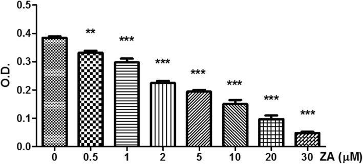
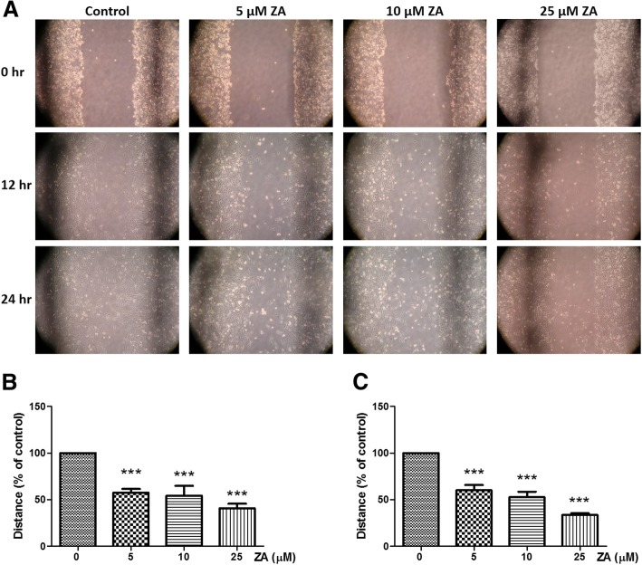
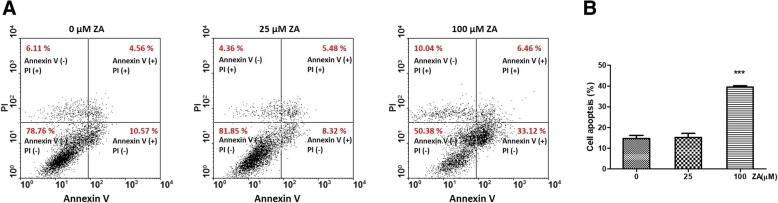
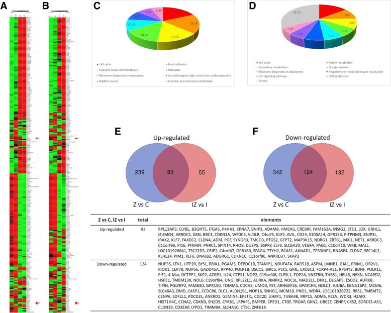
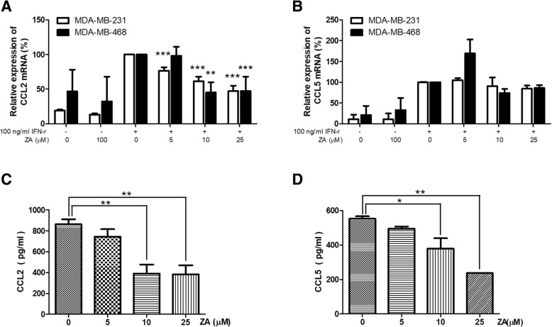
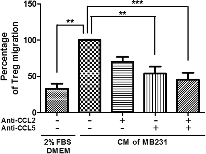
Methods: The anti-tumor effect of ZA on triple negative breast cancer cell lines were validated by XTT, wound healing and apoptosis analysis. A flow cytometry-based assay was used to analyze the immunosuppressive effect of Tregs treated with media conditioned by breast cancer cells, and a transwell assay was used to evaluate the chemotactic migration of Tregs. Differential gene expression profile on MDA-MB-231 treated with ZA (25 μM) was analyzed by. microarrays to describe the molecular basis of actions of ZA for possible direct anti-tumor effects. Enzyme-linked immunosorbent assays and quantitative real-time PCR were used to investigate the effect of ZA on the expression of cytokines/factors by breast cancer cells.
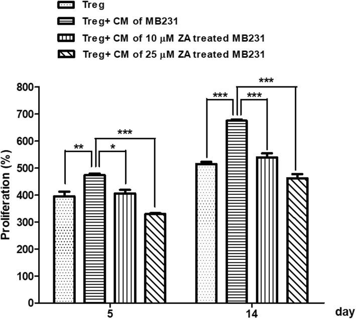
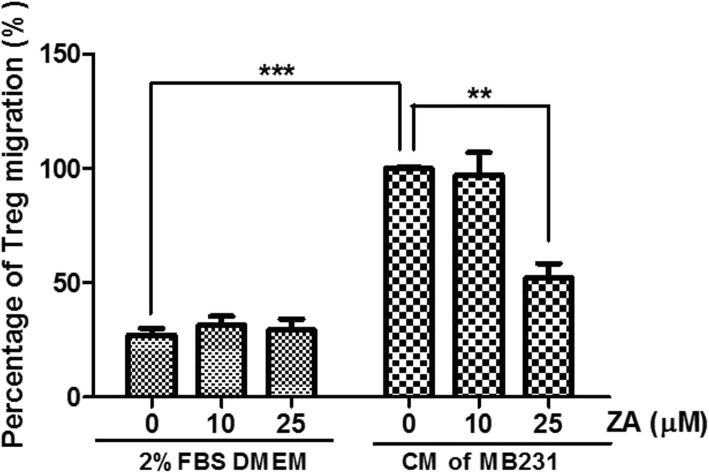
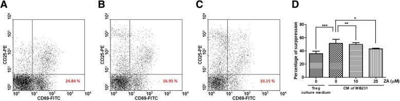
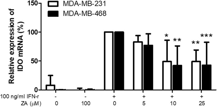
Results: ZA was found to inhibit the proliferation and migration of breast cancer cells. Media conditioned by the MDA-MB-231 cells promoted the expansion, chemotactic migration, and immunosuppressive activity of Tregs, and these effects were attenuated in a dose-dependent manner by ZA treatment, and the attenuation was due to reduced expression of selected breast cancer cell factors (CCL2, CCL5, and IDO).
Conclusions: ZA can significantly affect the interaction between breast cancer cells and Tregs. Our findings indicate that ZA is a potential therapeutic agent that can be used to reduce cancer aggressiveness by abolishing the supportive role of Tregs.
Keywords: Breast cancer; Immunomodulation; Regulatory T-cells; Zoledronic acid.
Conflict of interest statement
Ethics approval and consent to participate
The study was approved by the Institutional Review Committee of E-DA Hospital, and the volunteer donors of Treg cells provided written informed consent.
Consent for publication
Not applicable
Competing interests
The authors declare that they have no competing interests.
Publisher’s Note
Springer Nature remains neutral with regard to jurisdictional claims in published maps and institutional affiliations.
Figures
Similar articles
-
Immune modulation of CD4+CD25+ regulatory T cells by zoledronic acid.BMC Immunol. 2016 Nov 25;17(1):45. doi: 10.1186/s12865-016-0183-7. BMC Immunol. 2016. PMID: 27887569 Free PMC article.
-
Synergistic suppression of human breast cancer cells by combination of plumbagin and zoledronic acid In vitro.Acta Pharmacol Sin. 2015 Sep;36(9):1085-98. doi: 10.1038/aps.2015.42. Epub 2015 Aug 3. Acta Pharmacol Sin. 2015. PMID: 26235741 Free PMC article.
-
Zoledronic acid overcomes chemoresistance by sensitizing cancer stem cells to apoptosis.Biotech Histochem. 2018;93(2):77-88. doi: 10.1080/10520295.2017.1387286. Epub 2018 Jan 4. Biotech Histochem. 2018. PMID: 29300112
-
IDO, PTEN-expressing Tregs and control of antigen-presentation in the murine tumor microenvironment.Cancer Immunol Immunother. 2017 Aug;66(8):1049-1058. doi: 10.1007/s00262-017-2010-2. Epub 2017 May 9. Cancer Immunol Immunother. 2017. PMID: 28488123 Free PMC article. Review.
-
Anti-regulatory T cells.Semin Immunopathol. 2017 Apr;39(3):317-326. doi: 10.1007/s00281-016-0593-x. Epub 2016 Sep 27. Semin Immunopathol. 2017. PMID: 27677755 Review.
Cited by
-
The Case of Medication-Related Osteonecrosis of the Jaw Addressed from a Pathogenic Point of View. Innovative Therapeutic Strategies: Focus on the Most Recent Discoveries on Oral Mesenchymal Stem Cell-Derived Exosomes.Pharmaceuticals (Basel). 2020 Nov 25;13(12):423. doi: 10.3390/ph13120423. Pharmaceuticals (Basel). 2020. PMID: 33255626 Free PMC article. Review.
-
Targeting SREBP-2-Regulated Mevalonate Metabolism for Cancer Therapy.Front Oncol. 2020 Aug 21;10:1510. doi: 10.3389/fonc.2020.01510. eCollection 2020. Front Oncol. 2020. PMID: 32974183 Free PMC article. Review.
-
Zoledronic Acid Targeting of the Mevalonate Pathway Causes Reduced Cell Recruitment and Attenuates Pulmonary Fibrosis.Front Pharmacol. 2022 Jun 2;13:899469. doi: 10.3389/fphar.2022.899469. eCollection 2022. Front Pharmacol. 2022. PMID: 35721132 Free PMC article.
-
How zoledronic acid improves osteoporosis by acting on osteoclasts.Front Pharmacol. 2022 Aug 25;13:961941. doi: 10.3389/fphar.2022.961941. eCollection 2022. Front Pharmacol. 2022. PMID: 36091799 Free PMC article. Review.
-
Select HDAC Inhibitors Enhance Osteolysis and Bone Metastasis Outgrowth but Can Be Mitigated With Bisphosphonate Therapy.JBMR Plus. 2023 Jan 25;7(3):e10694. doi: 10.1002/jbm4.10694. eCollection 2023 Mar. JBMR Plus. 2023. PMID: 36936362 Free PMC article.
References
-
- Liu C, Workman CJ, Vignali DA. Targeting regulatory T cells in tumors. FEBS J. 2016;283(14):2731–2748. - PubMed
-
- Salama P, Phillips M, Grieu F, Morris M, Zeps N, Joseph D, Platell C, Iacopetta B. Tumor-infiltrating FOXP3+ T regulatory cells show strong prognostic significance in colorectal cancer. J Clin Oncol. 2009;27(2):186–192. - PubMed
-
- Chang LY, Lin YC, Mahalingam J, Huang CT, Chen TW, Kang CW, Peng HM, Chu YY, Chiang JM, Dutta A, et al. Tumor-derived chemokine CCL5 enhances TGF-beta-mediated killing of CD8(+) T cells in colon cancer by T-regulatory cells. Cancer Res. 2012;72(5):1092–1102. - PubMed
MeSH terms
Substances
Grants and funding
LinkOut - more resources
Full Text Sources
Research Materials
Miscellaneous
