. 2019 Apr 19;294(16):6494-6505.
doi: 10.1074/jbc.RA118.006071. Epub 2019 Feb 26.

Differential interactions of missing in metastasis and insulin receptor tyrosine kinase substrate with RAB proteins in the endocytosis of CXCR4

Affiliations

Differential interactions of missing in metastasis and insulin receptor tyrosine kinase substrate with RAB proteins in the endocytosis of CXCR4

Lushen Li et al. J Biol Chem. .

Abstract

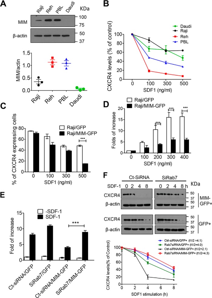
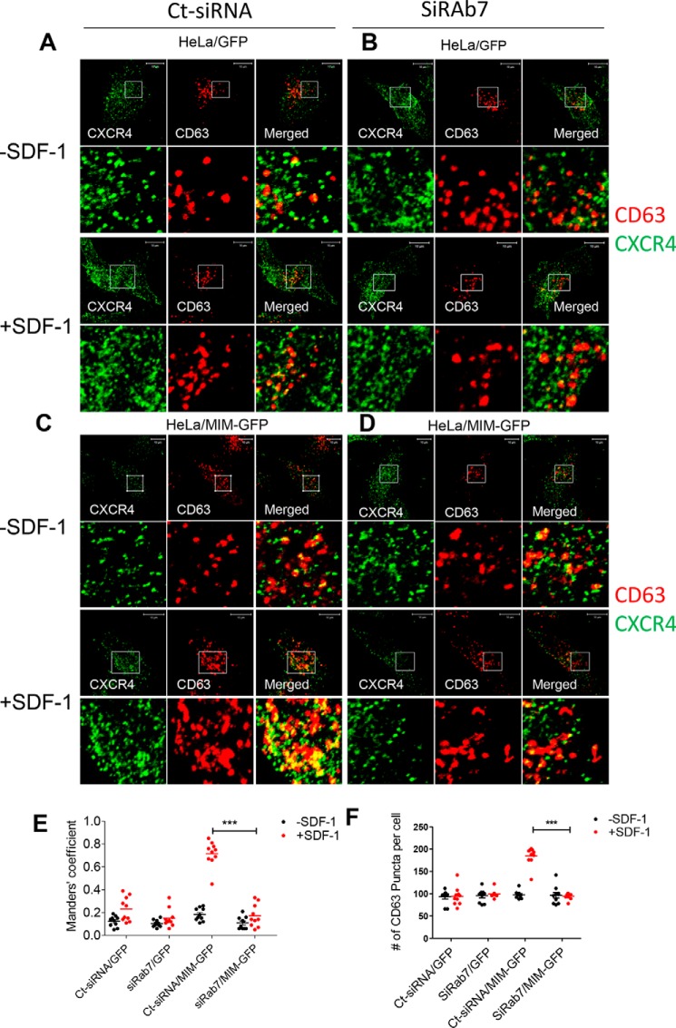
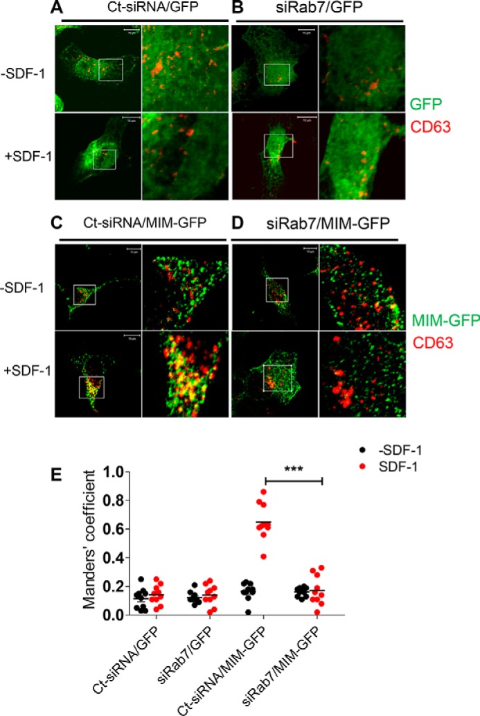
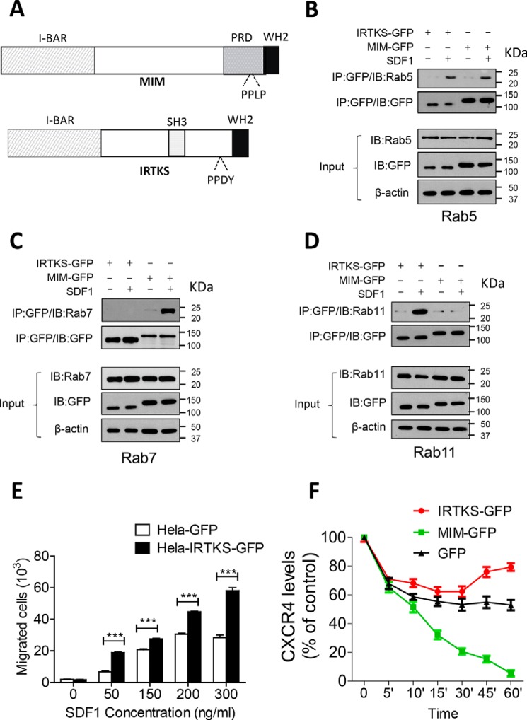
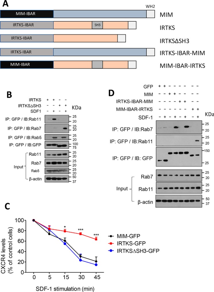
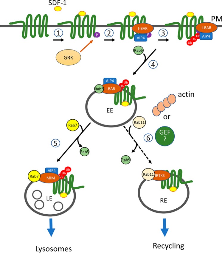
Missing in metastasis (MIM), an inverse Bin-Amphiphysin-Rvs (I-BAR) domain protein, promotes endocytosis of C-X-C chemokine receptor 4 (CXCR4) in mammalian cells. In response to the CXCR4 ligand stromal cell-derived factor 1 (SDF-1 or CXCL12), MIM associates with RAS-related GTP-binding protein 7 (RAB7) 30 min after stimulation. However, RAB7's role in MIM function remains undefined. Here we show that RNAi-mediated suppression of RAB7 expression in human HeLa cells has little effect on the binding of MIM to RAB5 and on the recruitment of CXCR4 to early endosomes but effectively abolishes MIM-mediated CXCR4 degradation, chemotactic response, and sorting into late endosomes and lysosomes. To determine whether I-BAR domain proteins interact with RAB7, we examined cells expressing insulin receptor tyrosine kinase substrate (IRTKS), an I-BAR domain protein bearing an Src homology 3 (SH3) domain. We observed that both MIM and IRTKS interact with RAB5 at an early response to SDF-1 and that IRTKS binds poorly to RAB7 but strongly to RAB11 at a later time point. Moreover, IRTKS overexpression reduced CXCR4 internalization and enhanced the chemotactic response to SDF-1. Interestingly, deletion of the SH3 domain in IRTKS abolished the IRTKS-RAB11 interaction and promoted CXCR4 degradation. Furthermore, the SH3 domain was required for selective targeting of MIM-IRTKS fusion proteins by both RAB7 and RAB11. Hence, to the best of our knowledge, our results provide first evidence that the SH3 domain is critical in the regulation of specific endocytic pathways by I-BAR domain proteins.

Keywords: C-X-C chemokine receptor type 4 (CXCR-4); RAB11; RAB7; Rab; SH3; cell signaling; chemokine; chemotaxis; endocytosis; endosome; insulin receptor tyrosine kinase substrate (IRTKS); missing in metastasis (MIM).


Conflict of interest statement

The authors declare that they have no conflicts of interest with the contents of this article. The content is solely the responsibility of the authors and does not necessarily represent the official views of the National Institutes of Health

Figures

Figure 1.
Figure 1.
RAB7 is required for MIM-mediated CXCR4 internalization. A, immunoblot analysis of endogenous MIM protein in Raji, Reh, Daudi, and PBL cells. The expression levels were quantified based on the ratio of MIM to β-actin. B, malignant B cells were stimulated with SDF-1 at the indicated concentrations for 30 min. Surface expression of CXCR4 was analyzed by flow cytometry and normalized to that of control cells without SDF-1 treatment. C, Raji cells were transiently transfected with GFP or MIM-GFP and stimulated for 30 min with SDF-1 at different concentrations. The percentage of cells expressing surface CXCR4 was determined by flow cytometry. D, Raji cells expressing MIM-GFP or GFP were plated on Transwell plates in which the lower chamber was filled with medium containing SDF-1 at the indicated concentrations. After 24 h, cells were fixed and stained with 0.1% crystal violet. The number of cells that migrated to the lower chamber was compared with that of cells without SDF-1 treatment. E, HeLa cells expressing GFP or MIM-GFP were treated with siRAB7 or Ct-siRNA for 48 h. The treated cells were then subjected to a Transwell assay for their chemotactic response to 500 ng/ml SDF-1. F, HeLa cells expressing either MIM-GFP or GFP were treated with siRAB7 or Ct-SiRNA. The treated cells were further incubated with 500 μg/ml cycloheximide for 30 min prior to exposure to 150 ng/ml SDF-1 for the indicated times. The total amounts of CXCR4 protein in treated cells at different times were estimated by immunoblot and used to calculate t½ using Prism software. All data represent mean ± S.E.M. (n = 3). ***, p < 0.001.
Figure 2.
Figure 2.
RAB7 is essential for MIM to promote CXCR4 sorting into late endosomes. A–D, HeLa cells expressing GFP (A and B) or MIM-GFP (C and D) were plated in 6-well plates, treated with Ct-siRNA (A and C) or siRAB7 (B and D) for 48 h, and stimulated with 500 ng/ml SDF-1 for 30 min. The treated cells were costained with monoclonal CXCR4 antibody (green) and polyclonal CD63 antibody (red) and inspected by confocal microscopy. The boxed areas of images were amplified and are presented below. E, co-localization of CXCR4 and CD63 was quantitively analyzed based on Manders' coefficient, which represents the proportion of CD63 colocalized with CXCR4. F, the amount of CD63 puncta of the acquired images was also quantified. Scale bars = 10 μm. ***, p < 0.001 (n = 10).
Figure 3.
Figure 3.
RAB7 is indispensable for recruitment of MIM into late endosomes. A–D, HeLa cells expressing GFP (A and B) or MIM-GFP (C and D) were treated with control siRNA (A and C) or siRAB7 (B and D) for 2 days prior to treatment with 500 ng/ml SDF-1 for 30 min. The treated cells were co-stained with GFP antibody (green) and CD63 antibody (red) and inspected by confocal microscopy. The boxed areas were amplified and are presented to the left of the corresponding images. E, co-localization of CD63 and GFP or MIM-GFP was quantified as described in the legend of Fig. 4. Data represent mean. ***, p < 0.001 (n = 10). Scale bars = 10 μm.
Figure 4.
Figure 4.
MIM and IRTKS were targeted by different Rabs. A, schematic of the MIM and IRTKS proteins. WH2, WASP homology 2; PRD, proline-rich domain; PPLP, proline-proline-leucine-proline residue sequence. B–D, HeLa cells expressing MIM-GFP or IRTKS-GFP were treated with 150 ng/ml SDF-1 for 5 min (B) or 30 min (C and D). Interactions between I-BAR domain proteins with RAB5 (B), RAB7 (C), and RAB11 (D) were analyzed by co-immunoprecipitation (IP), followed by immunoblot (IB) with the indicated antibodies. E, HeLa cells expressing GFP or IRTKS-GFP were plated in Transwell plates, treated with SDF-1 at different concentrations, and analyzed for chemotaxis as described in the legend of Fig. 1D. F, HeLa cells expressing GFP, MIM-GFP, and IRTKS-GFP were incubated with 500 μg/ml cycloheximide for 30 min prior to treatment with 150 ng/ml SDF-1 for the indicated times. The levels of CXCR4 on the surface of treated cells were estimated by flow cytometry. All data represent mean ± S.E.M. (n = 3). ***, p < 0.001
Figure 5.
Figure 5.
The SH3 domain determines recruitment of IRTKS to RAB11. A, schematic of MIM, IRTKS, and their fusion mutants. WH2, WASP homology 2. B, HeLa cells expressing GFP-tagged IRTKS or IRTKSΔSH3 proteins were stimulated with 150 ng/ml SDF-1 for 30 min. The cell lysates were subjected to immunoprecipitation (IP) with GFP antibody, followed by immunoblot (IB) using anti-RAB11, RAB5, or RAB7, as indicated. C, cells expressing GFP-tagged MIM, IRTKS, and IRTKSΔSH3 were treated with 500 ng/ml SDF-1 for up to 45 min. The levels of surface CXCR4 were estimated by flow cytometry. The data represent mean ± S.E.M. (n = 3). *, p < 0.05; ***, p < 0.001; referring to the differences between cells expressing IRTKS-GFP and those expressing IRTKSΔSH3-GFP. D, cells expressing MIM-GFP or its fusions were treated with 150 ng/ml SDF-1 for 30 min. The interactions of MIM and its fusions with RAB7 and RAB11 were analyzed as described in B.
Figure 6.
Figure 6.
A model depicting the function of I-BAR domain proteins in the regulation of CXCR4. CXCR4 is a G protein–coupled receptor with seven transmembrane domains. Upon SDF-1 stimulation, CXCR4 is phosphorylated at its C terminus by a G protein–coupled receptor kinase (GRK) (1). The phosphorylated CXCR4 allows its association with the E3 ubiquitin ligase AIP4, which is promoted by the I-BAR domain protein MIM or IRTKS. This results in formation of a complex of the CXCR4, AIP4, and I-BAR proteins (2) and a subsequent increase in ubiquitination of the receptor (3). Because of the ability to bind to RAB5 and RAB7, MIM facilitates sorting of the ubiquitinated receptor complex from early endosomes (EE) to late endosomes (LE) (5). On the other hand, IRTKS facilitates sorting of the receptor complex into a type of RAB11-coated endocytic vesicles, such as recycling vesicles (RE) (6). This step may be facilitated by actin assembly or an unidentified GEF of RAB11.

References

    1. Xia S., Li X., Johnson T., Seidel C., Wallace D. P., and Li R. (2010) Polycystin-dependent fluid flow sensing targets histone deacetylase 5 to prevent the development of renal cysts. Development 137, 1075–1084 10.1242/dev.049437 - DOI - PMC - PubMed
    1. Saarikangas J., Mattila P. K., Varjosalo M., Bovellan M., Hakanen J., Calzada-Wack J., Tost M., Jennen L., Rathkolb B., Hans W., Horsch M., Hyvönen M. E., Perälä N., Fuchs H., Gailus-Durner V., et al. (2011) Missing-in-metastasis MIM/MTSS1 promotes actin assembly at intercellular junctions and is required for integrity of kidney epithelia. J. Cell Sci. 124, 1245–1255 10.1242/jcs.082610 - DOI - PubMed
    1. Saarikangas J., Kourdougli N., Senju Y., Chazal G., Segerstrale M., Minkeviciene R., Kuurne J., Mattila P. K., Garrett L., Hölter S. M., Becker L., Racz I., Hans W., Klopstock T., Wurst W., et al. (2015) MIM-induced membrane bending promotes dendritic spine initiation. Dev. Cell 33, 644–659 10.1016/j.devcel.2015.04.014 - DOI - PubMed
    1. Yu D., Zhan X. H., Zhao X. F., Williams M. S., Carey G. B., Smith E., Scott D., Zhu J., Guo Y., Cherukuri S., Civin C. I., and Zhan X. (2012) Mice deficient in MIM expression are predisposed to lymphomagenesis. Oncogene 31, 3561–3568 10.1038/onc.2011.509 - DOI - PMC - PubMed
    1. Brown A. S., Meera P., Altindag B., Chopra R., Perkins E. M., Paul S., Scoles D. R., Tarapore E., Magri J., Huang H., Jackson M., Shakkottai V. G., Otis T. S., Pulst S. M., Atwood S. X., and Oro A. E. (2018) MTSS1/Src family kinase dysregulation underlies multiple inherited ataxias. Proc. Natl. Acad. Sci. U.S.A. 115, E12407–E12416 10.1073/pnas.1816177115 - DOI - PMC - PubMed

Publication types

MeSH terms

LinkOut - more resources