. 2019 Feb 26;116(9):3572-3577.
doi: 10.1073/pnas.1814526116. Epub 2019 Feb 11.

Snapshot of an oxygen intermediate in the catalytic reaction of cytochrome c oxidase

Affiliations

Snapshot of an oxygen intermediate in the catalytic reaction of cytochrome c oxidase

Izumi Ishigami et al. Proc Natl Acad Sci U S A. .

Abstract

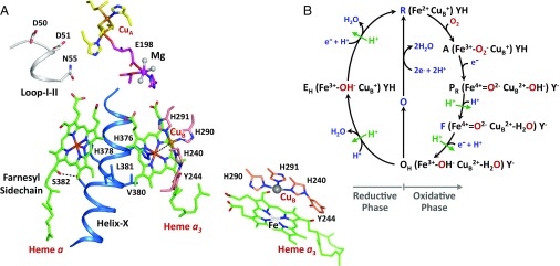
Cytochrome c oxidase (CcO) reduces dioxygen to water and harnesses the chemical energy to drive proton translocation across the inner mitochondrial membrane by an unresolved mechanism. By using time-resolved serial femtosecond crystallography, we identified a key oxygen intermediate of bovine CcO. It is assigned to the PR-intermediate, which is characterized by specific redox states of the metal centers and a distinct protein conformation. The heme a3 iron atom is in a ferryl (Fe4+ = O2-) configuration, and heme a and CuB are oxidized while CuA is reduced. A Helix-X segment is poised in an open conformational state; the heme a farnesyl sidechain is H-bonded to S382, and loop-I-II adopts a distinct structure. These data offer insights into the mechanism by which the oxygen chemistry is coupled to unidirectional proton translocation.

Keywords: X-ray free electron laser; bioenergetics; catalytic intermediates; complex IV; crystallography.


Conflict of interest statement

The authors declare no conflict of interest.

Figures

Fig. 1.
Fig. 1.
Active site structure of bCcO (A) and the proposed dioxygen reduction reaction cycle (B). The structure shown in A illustrates the relative location of the four redox centers, CuA, heme a and the CuB-heme a3 binuclear center, as well as the Mg site intervening the CuA center and the binuclear center. Helix-X (labeled in blue) contains the axial ligands of heme a3 and heme a, H376 and H378, respectively. (Lower Right Inset) Expanded view of the binuclear center. One of the CuB ligands, H240, is posttranslationally linked to Y244. All these structural elements are in subunit I, except CuA and E198(II) reside in subunit II. (B) Electronic configuration of the heme a3 iron, CuB, and Y244 in each intermediate is depicted. (See Introduction for the full description of the reaction sequence.) Each green arrow indicates the translocation of one pumped proton coupled to the designated reaction step (6).
Fig. 2.
Fig. 2.
Design and evaluation of the hydrodynamic focusing mixer (A and B) and time-resolved spectra associated with in crystallo dioxygen reduction reaction of bCcO (C and D). The schematic drawing in A illustrates the design of the mixer assembly. (B) Mixing behavior of the mixer simulated with COMSOL software, based on a convection-diffusion model. The two side streams, each flowing at a rate of 16 μL/min, squeeze the oxygen-containing central stream, flowing at a rate of 8 μL/min, such that complete mixing takes place before the mixed solution exits the mixer. The color bar indicates the normalized concentration of oxygen. (C) Microscopic image of typical bCcO microcrystals. (D) Optical absorption spectrum of the intermediate populated at 8 s after the mixing of a reduced bCcO microcrystal solution with an O2-containing buffer. The spectra of the reduced and oxidized forms of the enzyme are shown as references.
Fig. 3.
Fig. 3.
Structural changes associated with the R → PR transition. The structure of the PR-intermediate (green) is superimposed on reduced bCcO (R, coral). (Insets) Expanded views of the structural regions of (i) E198(II), highlighting the resemblance of the C-O-Mg angle in PR vs. R, demonstrating that CuA is in its reduced state; (ii) Loop-I-II, highlighting the large-scale movement of D51 and N55; and (iii) the [380-384] segment of Helix-X, showing the partial opening of the helical structure and the rotation in R438. The black curved arrows indicate RPR structural changes. The coral and green dashed lines represent H-bonds in reduced and intermediate structures, respectively.
Fig. 4.
Fig. 4.
Ligand density in the binuclear center of the PR-intermediate of bCcO. A shows the 2Fo-Fc electron density map of the initial reduced bCcO (R), demonstrating that no ligand density is present in its binuclear center. (B) Fo-Fc difference density map of the PR-intermediate, obtained without any ligand modeled, indicating the presence of exogenous ligand or ligands in the binuclear center. (C and D) Structure of the PR-intermediate modeled with a ferryl oxygen coordinated to the heme a3 and a hydroxide coordinated to CuB. (C) Polder OMIT map for each ligand. (D) 2Fo-Fc map. The CuB to heme a3 iron distance (in Å) is indicated in each structure. The Fo-Fc density map (green) is contoured at σ = 3, the Polder map (black) is contoured at σ = 7, and the 2Fo-Fc density maps (blue) are contoured at σ = 2.
Fig. 5.
Fig. 5.
A water molecule detected in the binuclear center of the PR-intermediate. The water molecule (W1) resides at the end of the K-channel, which may stabilize the tyrosinate configuration of Y244 and support proton transfer to the binuclear center. The numbers indicate the distances (in Å). The conserved water molecule, W2, is shown as a reference. The 2Fo-Fc difference map is contoured at σ = 1.
Fig. 6.
Fig. 6.
Postulated heme a gating mechanism. The mechanism is based on conformational changes induced by the change in the oxidation state of heme a and the associated rotation of the farnesyl side chain, as described in the text. PLS, proton loading site.

Similar articles

Cited by

References

    1. Yoshikawa S, Shimada A. Reaction mechanism of cytochrome c oxidase. Chem Rev. 2015;115:1936–1989. - PubMed
    1. Belevich I, Verkhovsky MI. Molecular mechanism of proton translocation by cytochrome c oxidase. Antioxid Redox Signal. 2008;10:1–29. - PubMed
    1. Han S, Takahashi S, Rousseau DL. Time dependence of the catalytic intermediates in cytochrome c oxidase. J Biol Chem. 2000;275:1910–1919. - PubMed
    1. Morgan JE, Verkhovsky MI, Palmer G, Wikström M. Role of the PR intermediate in the reaction of cytochrome c oxidase with O2. Biochemistry. 2001;40:6882–6892. - PubMed
    1. Morgan JE, Li PM, Jang DJ, el-Sayed MA, Chan SI. Electron transfer between cytochrome a and copper A in cytochrome c oxidase: A perturbed equilibrium study. Biochemistry. 1989;28:6975–6983. - PubMed

Publication types