Review
. 2019 Feb 26;12(570):eaav5183.
doi: 10.1126/scisignal.aav5183.

Specificity, versatility, and control of TGF-β family signaling

Affiliations
Review

Specificity, versatility, and control of TGF-β family signaling

Rik Derynck et al. Sci Signal. .

Abstract

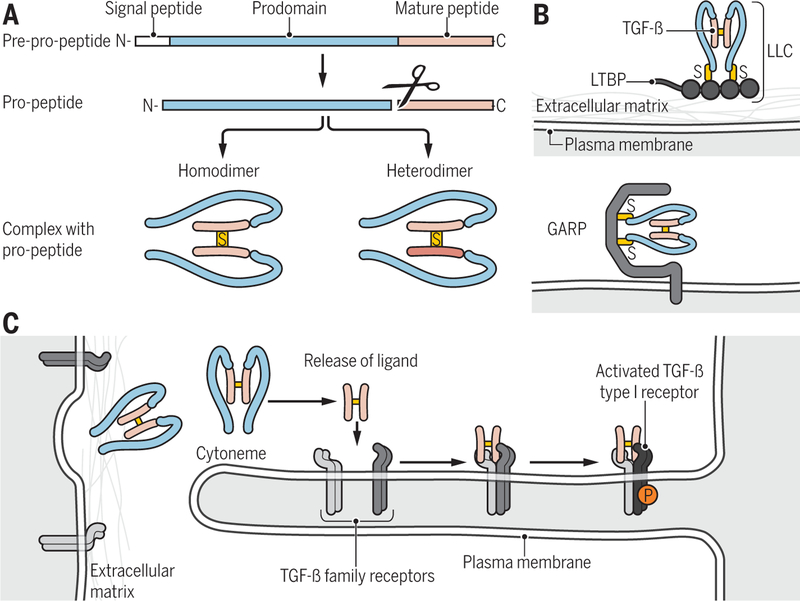
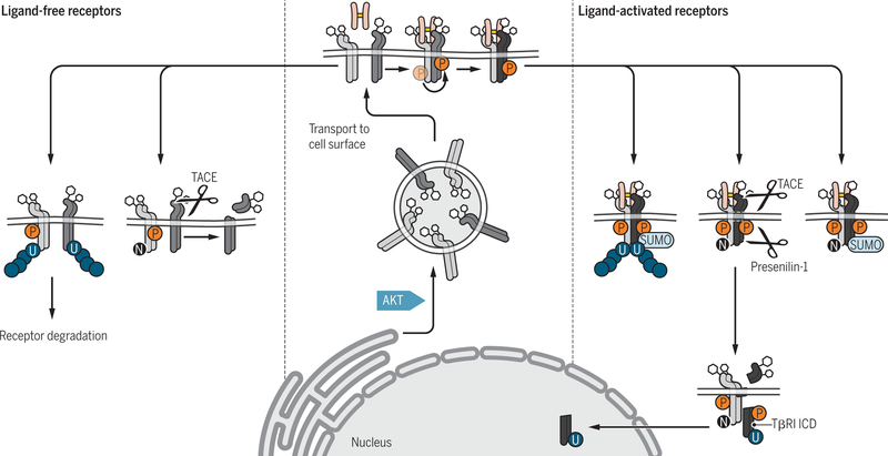
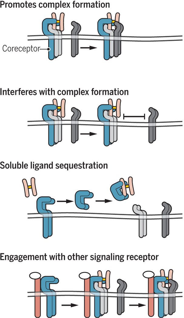
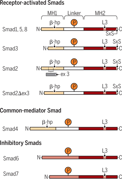
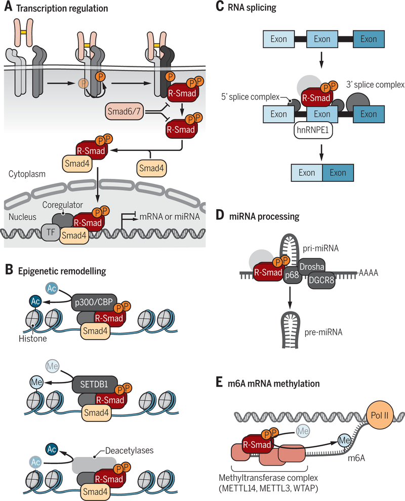
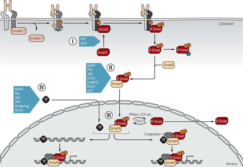
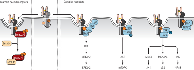
Encoded in mammalian cells by 33 genes, the transforming growth factor-β (TGF-β) family of secreted, homodimeric and heterodimeric proteins controls the differentiation of most, if not all, cell lineages and many aspects of cell and tissue physiology in multicellular eukaryotes. Deregulation of TGF-β family signaling leads to developmental anomalies and disease, whereas enhanced TGF-β signaling contributes to cancer and fibrosis. Here, we review the fundamentals of the signaling mechanisms that are initiated upon TGF-β ligand binding to its cell surface receptors and the dependence of the signaling responses on input from and cooperation with other signaling pathways. We discuss how cells exquisitely control the functional presentation and activation of heteromeric receptor complexes of transmembrane, dual-specificity kinases and, thus, define their context-dependent responsiveness to ligands. We also introduce the mechanisms through which proteins called Smads act as intracellular effectors of ligand-induced gene expression responses and show that the specificity and impressive versatility of Smad signaling depend on cross-talk from other pathways. Last, we discuss how non-Smad signaling mechanisms, initiated by distinct ligand-activated receptor complexes, complement Smad signaling and thus contribute to cellular responses.


Figures

Figure 1.
Figure 1.. Ligand processing and presentation.
(A) TGF-β family proteins are synthesized as precursor molecules consisting of a signal peptide, a prodomain (termed latency-associated peptide, LAP, for TGF-β), and the mature polypeptide. After signal peptide removal, the precursor is further processed by proteolytic cleavage at basic residues, thus separating the prodomain from the mature polypeptide, which remain non-covalently associated. Concomitant, disulfide-linked dimerization of the mature polypeptides into mature homo- and heterodimeric proteins is shown. (B) Latent TGF-β complex can associate through disulfide bonding with LTBP into a large latent complex (LLC) that in turn associates with the extracellular matrix (top), or with the plasma membrane-associated GARP (bottom). (C) Cytoneme-associated activation of TGF-β family signaling. Long cytonemes extend from the cell body, and present TGF-β family receptors to ligand complexes. Binding of ligand to the receptors results in activation of the type I receptors (light to dark blue). Credit: Veronica Falconieri Hays/Science Signaling
Figure 2.
Figure 2.. Posttranslational and functional modifications of the glycosylated TGF-β receptors TβRII and TβRI before or after ligand-induced activation.
AKT activation in response to insulin or other stimuli drives TGF-β receptor transport from intracellular compartments to the cell surface (center). Prior to activation (left, light blue), the plasma membrane-associated TβRI (blue) can undergo TACE-mediated ectodomain cleavage, thus preventing ligand-induced activation of signaling, or polyubiquitylation that leads to receptor degradation. Ligand-induced activation of the TβRI receptor (right, dark blue) results in phosphorylation (P) of its GS domain by TβRII (green), and phosphorylation of TβRII and TβRI lead to TβRII neddylation (N), TβRI sumoylation (SUMO), and TβRII and TβRI ubiquitylation (U). Activation of the TβRI receptor may also result in proteolytic release of its intracellular cytoplasmic domain (ICD) by presenilin-1 after TACE-mediated ectodomain cleavage, and then nuclear translocation of the ICD. Credit: Veronica Falconieri Hays/Science Signaling
Figure 3.
Figure 3.. Roles of coreceptors in TGF-β family ligand binding to heteromeric complexes of type II (green) and type I receptors.
The membrane-anchored coreceptors (pink) are generally present as dimers at the cell surface and can promote ligand binding to the receptors (top left), as shown for betaglycan, which enhances binding of TGF-β2 to the TGF-β receptors, or for crypto which enables nodal binding to activin receptors, and nodal signaling. Alternatively, association of coreceptors with type II receptors (green) can interfere with the formation of complexes between type II and type I (blue) receptors (top right), as reported for the roles of RGMs as BMP coreceptors. Co-receptors, such as betaglycan and endoglin, can also be cleaved at the cell surface, resulting in the release of their ectodomains (bottom left). The released ectomains retain their affinity for ligand, resulting in ligand sequestration and repression of signaling activation. Co-receptors also provide opportunities to coordinate activation of distinct signaling pathways (bottom right). Thus, betaglycan can bind bFGF in addition to TGF-β and coordinately regulate FGF and TGF-β signaling. Credit: Veronica Falconieri Hays/Science Signaling
Figure 4.
Figure 4.. Schematic comparison of the simplified structures of R-Smads (Smad1, Smad2, Smad3, Smad5 and Smad8), Smad4, and inhibitory Smads (Smad6 and Smad7).
The R-Smads and Smad4 have two conserved domains, the MH1 (brown-grey) and MH2 (dark orange) domains, separated by a variable serine- and proline-rich linker region (light grey). The linker region is targeted for phosphorylation (P) by various signaling kinases that thus control the stabilities and functions of the Smads. A β-hairpin (β-hp; arrow head), which in Smad2 is interrupted by a sequence encoded by exon 3 but is maintained in the Smad2Δ3 variant, enables MH1 domain binding to DNA. The inhibitory Smads lack an MH1 domain, and have a long and variable sequence (light orange) preceding the MH2 domain. This sequence is thought to be structurally versatile depending on post-translational modifications and protein interactions. The positively charged L3 loop in the MH2 domain mediates association with the activated type I receptors and with other Smads. The R-Smads, but not Smad4 and the inhibitory Smads, have a conserved C-terminal SXS motif that is phosphorylated by the activated type I receptor, resulting in R-Smad activation. Credit: Veronica Falconieri Hays/Science Signaling
Figure 5.
Figure 5.. Smad-dependent regulation of gene expression.
(A) Simplified model of TGF-β-induced R-Smad activation leading to Smad-mediated activation of gene expression. Signaling is initiated by TGF-β binding to a heteromeric complex of type II (green) and type I (blue) receptors, resulting in activation of the type I receptors (dark blue) and C-terminal R-Smad phosphorylation. The activated R-Smads dissociate from the type I receptors, form a complex with Smad4 and the R-Smad/Smad4 complexes translocate into the nucleus, where they regulate gene expression with transcription factors (TF) and coregulators. Inhibitory Smads (Smad6 and Smad7) interfere with functional Smad activation, by associating with type I receptors, thus preventing R-Smad activation, or by interfering with the complex formation of R-Smads with Smad4. (B) Activated R-Smad/Smad4 complexes associate and cooperate with high affinity DNA-binding transcription factors to activate or repress the transcription of genes into mRNA or microRNA precursors. (C) Activated R-Smad/Smad4 complexes recruit histone modifying enzymes, resulting in chromatin remodeling. Recruitment of the p300 acetyltransferase, which commonly acts as transcription coactivator for Smad complexes, confers H3K9 acetylation, whereas recruitment of the SETDB1methyltransferase induces H3K9 methylation and thus represses transcription. Smad-mediated recruitment of histone deacetylases leads to histone deacetylation (not depicted). (D) TGF-β-activated Smad complexes regulate mRNA splicing in association with hnRNPE1. (E) Activated R-Smads direct miRNA processing through association with the p68 RNA helicase in complex with Drosha RNAse. (F) Activated Smad2 or Smad3 can associate with m6A methyltransferase complexes to promote methylation of mRNA. Credit: Veronica Falconieri Hays/Science Signaling
Figure 6.
Figure 6.. Signaling crosstalk through posttranslational control of Smad activation and functions.
R-Smad association with the receptors (I) is controlled by inhibitory Smad6 and/or Smad7, which prevent R-Smad access to the activated type I receptors (dark blue). Additionally, upon activation in response to various signaling pathways, AKT and IRF3 bind to Smad3 and thus attenuate Smad3 binding to activated type I receptors. Various signaling pathways that act through kinases target the linker regions of R-Smads for phosphorylation (II), with the possibility for further regulation by subsequent dephosphorylation, and thus control the subcellular localization, stability and function of Smads. Smads can also be poly-ubiquitylated, leading to degradation, and in some cases targeted linker phosphorylation is a prerequisite for subsequent poly-ubiquitylation and degradation. Some kinases and a phosphatase are listed as examples. In the nucleus (III), phosphorylation and dephosphorylation by kinases and phosphatases further regulate the Smad activities. Direct transcriptional activation or repression of target genes requires association of Smad complexes with DNA-binding transcription factors (TFs) and coregulators (IV). Smads have been shown to associate with a wide variety of TFs, depending on the signaling status of the cells and the targeted gene. Extensive signaling crosstalk occurs at the level of Smad-complex association with DNA-binding TFs, because they are also regulated by phosphorylation or other modifications in response to signaling pathways. Some examples are listed. Such crosstalk may occur before binding of Smad-TF complexes to regulatory gene sequences or after formation of the DNA-binding nucleoprotein complexes. Credit: Veronica Falconieri Hays/Science Signaling
Figure 7.
Figure 7.. TGF-β receptors activate Smad signaling and non-Smad signal transduction pathways.
Smad-mediated signaling occurs in association with nascent clathrin-dependent endosomal compartments, while receptor-induced non-Smad signaling pathways emanate from caveolar compartments or are not yet known to associate with either type of compartment. TGF-β-induced ERK/MAPK pathway activation occurs in caveolar lipid raft compartments and requires ShcA. TGF-β-induced PI3K-AKT signaling has also been shown to emanate from caveolar lipid raft compartments and to require ShcA, raising the possibility that both pathways initiate from the same receptor complexes. However, TGF-β-induced AKT activation was also shown in different cells to require TRAF6 and to not require the TβRI kinase activity, suggesting that it initiates from different receptor complexes, as shown. TGF-β-induced p38 MAPK and JNK activation has been shown to require TRAF6 and to be initiated by TAK1 activation, while TGF-β-induced p38 MAPK activation has been localized to cholesterol-rich lipid raft compartments, suggesting the existence of distinct complexes for TGF-β-induced p38MAPK, JNK and NFκB signaling, as shown. Whether Smad7 association with the type I receptor facilitates, is required for or antagonizes TGF-β-induced p38 MAPK activation is unclear because of seemingly conflicting reports. Credit: Veronica Falconieri Hays/Science Signaling

Similar articles

Cited by

References

    1. Derynck R, Miyazono K, The Biology of the TGF-β Family. (Cold Spring Harbor Laboratory Press, New York, ed. Derynck Rik, Miyazono Kohei 2016), pp. 1164.
    1. Morikawa M, Derynck R, Miyazono K, TGF-β and the TGF-β family: context-dependent roles in cell and tissue physiology. Cold Spring Harb Perspect Biol 8, a021873 (2016). - PMC - PubMed
    1. Moses HL, Roberts AB, Derynck R, The discovery and early days of TGF-β: a historical perspective. Cold Spring Harb Perspect Biol 8, a021865 (2016). - PMC - PubMed
    1. Derynck R, Jarrett JA, Chen EY, Eaton DH, Bell JR, Assoian RK, Roberts AB, Sporn MB, Goeddel DV, Human transforming growth factor-β complementary DNA sequence and expression in normal and transformed cells. Nature 316, 701–705 (1985). - PubMed
    1. Zhang Y, Alexander PB, Wang XF, TGF-β family signaling in the control of cell proliferation and survival. Cold Spring Harb Perspect Biol 9, a022145 (2017). - PMC - PubMed

Publication types

MeSH terms

Substances