. 2019 Feb 26;9(1):2846.
doi: 10.1038/s41598-018-37564-w.

Validation of vessel size imaging (VSI) in high-grade human gliomas using magnetic resonance imaging, image-guided biopsies, and quantitative immunohistochemistry

Affiliations

Validation of vessel size imaging (VSI) in high-grade human gliomas using magnetic resonance imaging, image-guided biopsies, and quantitative immunohistochemistry

Ararat Chakhoyan et al. Sci Rep. .

Abstract

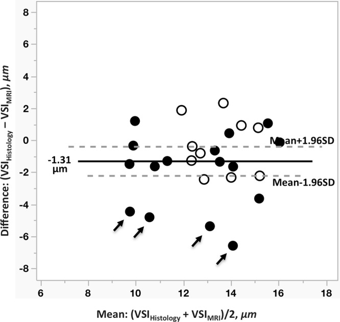
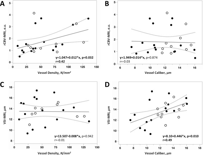
To evaluate the association between a vessel size index (VSIMRI) derived from dynamic susceptibility contrast (DSC) perfusion imaging using a custom spin-and-gradient echo echoplanar imaging (SAGE-EPI) sequence and quantitative estimates of vessel morphometry based on immunohistochemistry from image-guided biopsy samples. The current study evaluated both relative cerebral blood volume (rCBV) and VSIMRI in eleven patients with high-grade glioma (7 WHO grade III and 4 WHO grade IV). Following 26 MRI-guided glioma biopsies in these 11 patients, we evaluated tissue morphometry, including vessel density and average radius, using an automated procedure based on the endothelial cell marker CD31 to highlight tumor vasculature. Measures of rCBV and VSIMRI were then compared to histological measures. We demonstrate good agreement between VSI measured by MRI and histology; VSIMRI = 13.67 μm and VSIHistology = 12.60 μm, with slight overestimation of VSIMRI in grade III patients compared to histology. rCBV showed a moderate but significant correlation with vessel density (r = 0.42, p = 0.03), and a correlation was also observed between VSIMRI and VSIHistology (r = 0.49, p = 0.01). The current study supports the hypothesis that vessel size measures using MRI accurately reflect vessel caliber within high-grade gliomas, while traditional measures of rCBV are correlated with vessel density and not vessel caliber.


Conflict of interest statement

B.E.; Advisory Board – Hoffman La-Roche; Siemens; Nativis; Medicenna; MedQIA; Bristol Meyers Squibb; Imaging Endpoints; Agios. Paid Consultant – Nativis; MedQIA; Siemens; Hoffman La-Roche; Imaging Endpoints; Medicenna; Agios.

Figures

Figure 1
Figure 1
Two representative high-grade glioma patients are reported with their respective biopsy targets. T2w-FLAIR (upper) or post-contrast T1w (bottom) images were used for anatomical reference in tissue sampling in various tumor grades. ADC, rCBV and VSIMRI maps are shown for each patient. The first patient (upper) has a WHO grade III, Anaplastic Oligodendroglioma. The brain lesion is located in the left frontal lobe. The second patient (bottom) is a WHO Grade IV glioblastoma with a brain lesion located in the left parietal lobe. An elevated ADC and rCBV showed in both patients within tumor areas. Glioblastoma patient show an increased heterogeneity of vessel size within and around enhancing areas. Examples of tissue slides with stained CD31 positive vessels for each representative target. As observed with VSIMRI, in both grade III and IV, CD31 positive vessels are present with spatial heterogeneity in terms of density and morphometry.
Figure 2
Figure 2
Bland-Altman plot represents potential bias of agreement between these two techniques. A number of points display higher difference between VSIMRI and VSIHistology, especially in grade III (black arrows). Filled and unfilled circles presenting targets from grade III and grade IV, respectively.
Figure 3
Figure 3
Linear correlation was performed between rCBV, VSIMRI and their corresponding histology value; vessel density and vessel caliber (VSIHistology). Positive linear correlation was found between rCBV vs. vessel density (A, r = 0.42, p = 0.032) as well as between VSIMRI and vessel caliber (D, r = 0.49, p = 0.010). However, no direct relationship was observed between rCBV and vessel caliber (B). VSIMRI measures are independent of vessel density (C). The filled and unfilled circles presenting targets from grade III and grade IV, respectively.

Similar articles

Cited by

References

    1. Jain RK, et al. Angiogenesis in brain tumours. Nat. Rev. Neurosci. 2007;8:610–22. doi: 10.1038/nrn2175. - DOI - PubMed
    1. Batchelor TT, et al. Improved tumor oxygenation and survival in glioblastoma patients who show increased blood perfusion after cediranib and chemoradiation. Proc. Natl. Acad. Sci. 2013;110:19059–64. doi: 10.1073/pnas.1318022110. - DOI - PMC - PubMed
    1. Hardee ME, Zagzag D. Mechanisms of glioma-associated neovascularization. Am. J. Pathol. 2012;181:1126–1141. doi: 10.1016/j.ajpath.2012.06.030. - DOI - PMC - PubMed
    1. Das S, Marsden PA. Angiogenesis in Glioblastoma. N. Engl. J. Med. 2013;369:1561–1563. doi: 10.1056/NEJMcibr1309402. - DOI - PMC - PubMed
    1. Mahase S, et al. Hypoxia-mediated mechanisms associated with antiangiogenic treatment resistance in Glioblastomas. Am. J. Pathol. 2017;187:940–953. doi: 10.1016/j.ajpath.2017.01.010. - DOI - PMC - PubMed

Publication types

MeSH terms