. 2019 Feb 26;9(1):2821.
doi: 10.1038/s41598-019-39541-3.

Tlr2 Deficiency is Associated with Enhanced Elements of Neuronal Repair and Caspase 3 Activation Following Brain Ischemia

Affiliations

Tlr2 Deficiency is Associated with Enhanced Elements of Neuronal Repair and Caspase 3 Activation Following Brain Ischemia

Dunja Gorup et al. Sci Rep. .

Abstract

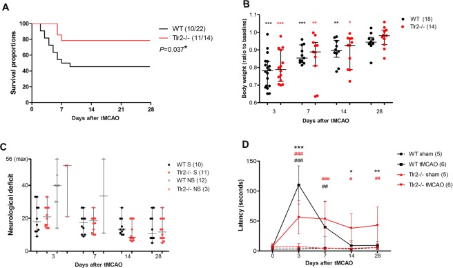
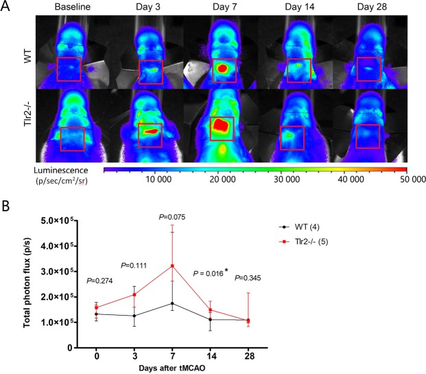
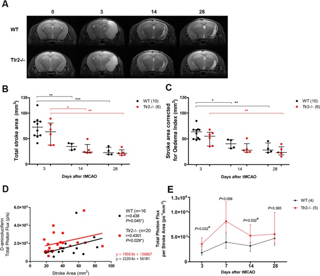
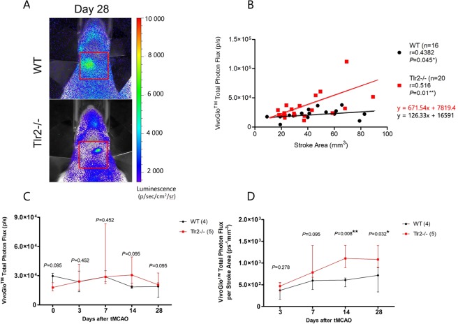
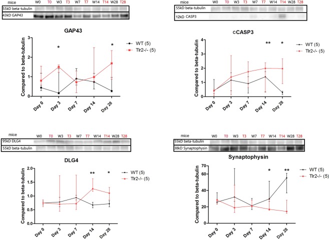
The aim of this study was to apply multimodal in vivo imaging to assess the influence of altered innate immunity on brain repair after ischemic lesion. Tlr2-deficient mice were compared to wild type controls, as they lack Tlr2-mediated pro-inflammatory signaling triggered by postischemic necrosis. The ischemic lesion was induced by transient middle cerebral artery occlusion for 60 min, followed by brain imaging and analysis at four time points until 28 days after ischemia. Multimodal in vivo imaging involved a combination of 3 modalities: (1) magnetic resonance imaging by T2-weighted scans to assess brain lesion size, (2) bioluminescence imaging of Gap43-luc/gfp transgenic mice to visualize the axonal remodeling, and (3) caged-luciferin bioluminescence imaging of DEVD-luciferin allowing for visualization of caspase 3 and 7 activity in Gap43-luc/gfp mice. This enabled innovative correlation of the MRI-determined lesion size to photon fluxes obtained by bioluminescence imaging. Our data revealed that following ischemia, Tlr2-deficient mice had higher Gap43 expression and higher levels of caspases 3 and 7 activity, which was accompanied by enhanced levels of synaptic plasticity markers DLG4 and synaptophysin when compared to wild type controls. Altered inflammation in Tlr2-deficient mice was accompanied by enhanced elements of post-stroke repair, in particular during the chronic phase of recovery, but also with delayed final consolidation of the brain lesion.


Conflict of interest statement

The authors declare no competing interests.

Figures

Figure 1
Figure 1
(A) Tlr2−/− mice survival after ischemic lesion is better than of their wild type (WT) counterparts analyzed by Log-rank (Mantel-Cox) test P = 0.037*; (B) Both, Tlr2−/− and WT lose body weight after ischemic lesion, and subsequently regain it after 28 days; (C) Mice that reached humane endpoint (grey and pale red) (non-survivors, NS) had worse neurological scores than their survivor counterparts. There was no significant difference between wild type (WT), and Tlr2−/− mice. (D) Bilateral tactile stimulation test revealed decreased latency in sticky tape removal from the contralateral forepaw in acute time points in Tlr2−/− mice compared to their wild type (WT) counterparts, but increased time of reaction in the later time points of 14 and 28 days. ***,** and *marking P < 0.001, P < 0.01, P < 0.05 for differences between WT controls and Tlr2−/− controls, respectively. #Are used to mark differences within the same genotype compared to their sham operated controls (WT controls vs. WT tMCAO, Tlr2−/− controls vs. Tlr2−/− tMCAO).
Figure 2
Figure 2
(A) Representative images of bioluminescence signal imaging using d-aminoluciferin for Gap43-luc-gfp (WT), and Gap43-luc-gfp-Tlr2−/− (Tlr2−/−) mice. (B) Total photon flux measured for Gap43-luc-gfp (WT), and Gap43-luc-gfp-Tlr2−/− (Tlr2−/−) mice showing median and interquartile range.
Figure 3
Figure 3
(A) Representative MRI T2 maps of ishemic lesion in WT and Tlr2−/− mice. (B) Total stroke area measured by a blinded rater shows gradual decrease in ischemic volume with no differences between the WT and Tlr2−/− mice. (C) Stroke area adjusted for oedema index shows the volume of ischemic lesion used for further calculation of correlations. (D) Correlations of BLI signal using d-aminoluciferin and Stroke area by genotype. (E) Total Photon Flux adjusted per stroke Area measured by MRI shows increased BLI signal in Tlr2−/− mice compared to their WT counterparts in acute and chronic time point (3 and 14 days).
Figure 4
Figure 4
(A) BLI imaging after VivoGloTM injection of representative Gap43-luc-gfp (WT), and Gap43-luc-gfp-Tlr2−/− (Tlr2−/−) mice; (B) VivoGloTM total photon flux in correlation with Stroke Area measured by MRI. (C) VivoGloTM total photon flux dynamics (D) VivoGloTM total photon flux adjusted to Stroke area measured by MRI indicating statistically significant increase of BLI signal for Tlr2−/− mice at Days 14 and 28.
Figure 5
Figure 5
Western Blot with analysis per time point of WT and TLR−/− showing median and interquartile ranges with statistically significant differences at chronic time points for GAP43 and CASP3, as well as for synaptic markers of DLG4 and synaptophysin. ***,** and *marking P < 0.001, P < 0.01, P < 0.05 for differences between WT controls and Tlr2−/− controls, respectively. The blots shown here were cut from the bigger gel, therefore boxed here, and the full-length blots were provided in Figure S1.

References

    1. Maiser SJ, et al. Intravenous recombinant tissue plasminogen activator administered after 3 h following onset of ischaemic stroke: a metaanalysis. International journal of stroke: official journal of the International Stroke Society. 2011;6:25–32. doi: 10.1111/j.1747-4949.2010.00537.x. - DOI - PubMed
    1. Indredavik B, Bakke F, Slordahl SA, Rokseth R, Haheim LL. Stroke unit treatment. 10-year follow-up. Stroke. 1999;30:1524–1527. doi: 10.1161/01.STR.30.8.1524. - DOI - PubMed
    1. Kahle MP, Bix GJ. Successfully Climbing the “STAIRs”: Surmounting Failed Translation of Experimental Ischemic Stroke Treatments. Stroke research and treatment. 2012;2012:374098. doi: 10.1155/2012/374098. - DOI - PMC - PubMed
    1. Bohacek I, et al. Toll-like receptor 2 deficiency leads to delayed exacerbation of ischemic injury. Journal of neuroinflammation. 2012;9:191. doi: 10.1186/1742-2094-9-191. - DOI - PMC - PubMed
    1. Lehnardt S, et al. Toll-like receptor 2 mediates CNS injury in focal cerebral ischemia. Journal of neuroimmunology. 2007;190:28–33. doi: 10.1016/j.jneuroim.2007.07.023. - DOI - PubMed

Publication types