. 2019 Feb 26;9(1):2836.
doi: 10.1038/s41598-019-39349-1.

Protective effect of oxytocin on LPS-induced acute lung injury in mice

Affiliations

Protective effect of oxytocin on LPS-induced acute lung injury in mice

Xiaona An et al. Sci Rep. .

Abstract

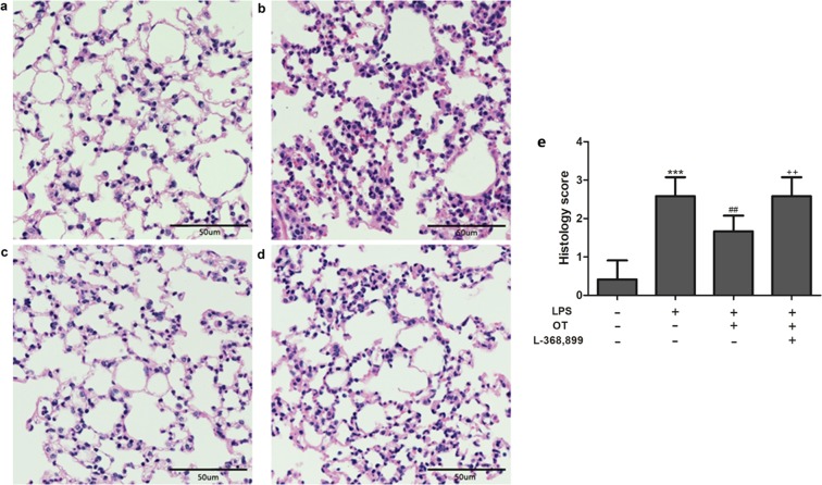
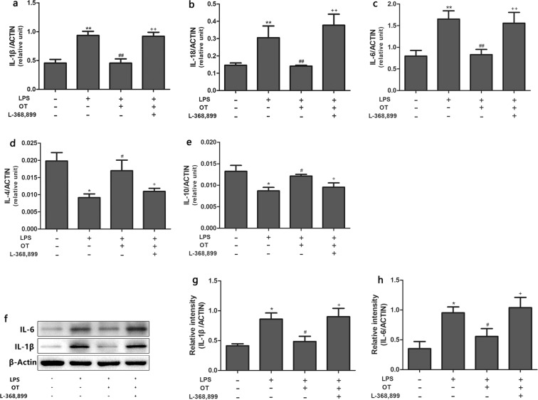
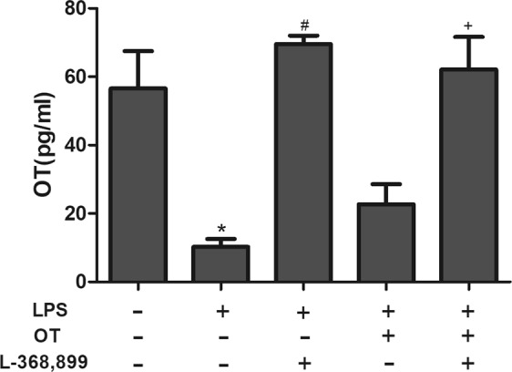
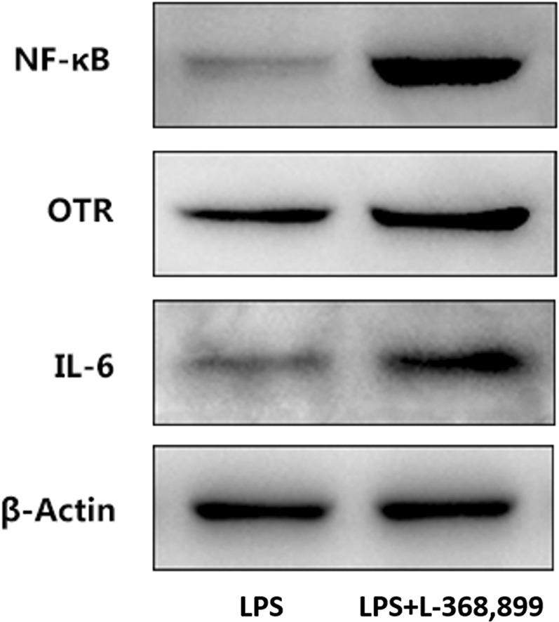
Oxytocin (OT), a neurohypophyseal hormone synthesized in the paraventricular and supraoptic nuclei of the hypothalamus, has been reported to have an anti- inflammatory effect. However, its role in acute lung injury (ALI) has never been investigated. The aim of this study was to explore the therapeutic effects and potential mechanism action of OT on lipopolysaccharide (LPS)-induced ALI. Mice were treated with OT 30 min before the intraperitoneal injection of LPS. After 2 h, the effects of OT on lung histopathological changes, lung wet/dry (W/D) ratio, myeloperoxidase (MPO) activity, levels of inflammatory cytokines in the bronchoalveolar lavage fluid (BALF), and expression of inflammation proteins were detected. The results showed that OT significantly reduced LPS-induced pathological injury, W/D ratio, MPO activity, and the levels of interleukin (IL)-1β, IL-18 and IL-6. Further, OT also inhibited LPS-induced Toll-like receptor 4 expression and NLR family pyrin domain containing 3 inflammasome activation. OT receptor antagonist (L-368,899) was given 90 min before injecting OT to further demonstrate the role of OT in LPS-induced ALI. The results showed OT could not alleviate the aforementioned inflammatory reactions after administering L-368,899. In conclusion, the present results indicated that OT could reduce inflammatory responses of LPS-induced ALI.


Conflict of interest statement

The authors declare no competing interests.

Figures

Figure 1
Figure 1
Effects of OT on the histological changes in lung tissue in mice with LPS-induced ALI. Representative histological changes in lungs obtained from mice of different groups. (a) Control group. (b) LPS group. (c) LPS + OT group. (d) LPS + OT + L-368,899 group. Pathological changes in lung tissues were observed using HE staining (light microscopy, ×200). The histopathologic scores are presented for the lung tissues (e). The data are presented as the means ± standard error of the mean. ***P < 0.001 vs control. ##P < 0.01 vs LPS. ++P < 0.01vs OT. Data show means with SEM for six mice/group.
Figure 2
Figure 2
Effects of OT on lung MPO activity in mice with LPS-induced ALI. The values are presented as means ± standard error of the mean. ***P < 0.001 vs control. ###P < 0.001 vs LPS. +++P < 0.001 vs OT. Data show means with SEM for five mice/group.
Figure 3
Figure 3
Effects of OT on the lung W/D ratio in mice with LPS-induced ALI. The values are presented as means ± standard error of the mean. **P < 0.01 vs control. ##P < 0.01 vs LPS. ++P < 0.01 vs OT. Data show means with C SEM for four mice/group.
Figure 4
Figure 4
Effects of OT on the production of inflammatory cytokines in BALF from mice with LPS-induced ALI. The levels of IL-1β (a), IL-18 (b), IL-6 (c), IL-4 (d), and IL-10 (e) were measured using ELISA. The values are presented as means ± standard error of the mean. **P < 0.01, ***P < 0.001 vs control. ##P < 0.01, ###P < 0.001vs LPS. ++P < 0.01, +++P < 0.001vs OT. Data show means with SEM for eight mice/group.
Figure 5
Figure 5
Effects of OT on the gene expression of inflammatory cytokines in the lung tissues from mice with LPS-induced ALI. The mRNA expression of IL-1β (a), IL-18 (b) IL-6 (c), IL-4 (d), and IL-10 (e) was measured using RT-qPCR. The proteins expression levels of IL-1β and IL-6 were analyzed using the Western blot analysis (f). The proteins expression of IL-1β and IL-6 were quantified by measuring band intensities and displayed as fold increase relative to ACTIN (g and h). The values are presented as means ± standard error of the mean. *P < 0.05, **P < 0.01 vs control. #P < 0.05, ##P < 0.01 vs LPS. +P < 0.05, ++P < 0.01 vs OT. Data show means with SEM for four to eight mice/group.
Figure 6
Figure 6
Effects of OT on the expression of OTR in the lung tissues from mice with LPS-induced ALI. The expression levels of OTR were evaluated using the Western blot analysis, IHC, and IF (a,b). The Western blot analysis, IHC, and IF of OTR were quantified respectively (ce). *P < 0.05, ***P < 0.001 vs control. #P < 0.05, ##P < 0.01, ###P < 0.001 vs LPS. +P < 0.05, +++P < 0.001 vs OT. Data show means with SEM for five mice/group.
Figure 7
Figure 7
Effects of OT on the expression of NF-κB and TLR4 in the lung tissues from mice with LPS-induced ALI. The expression levels of these proteins were evaluated using the Western blot analysis, IHC, and IF (a,d,g). The Western blot analysis, IHC, and IF of OTR were quantified respectively (b,c,e,f,h,i). *P < 0.05, **P < 0.001,***P < 0.001 vs control. #P < 0.05, ##P < 0.01, ###P < 0.001 vs LPS. +P < 0.05, +++P < 0.001 vs OT. Data show means with SEM for five mice/group.
Figure 8
Figure 8
Effects of OT on the expression of NLRP3 and Caspase-1 in the lung tissues from mice with LPS-induced ALI. The expression levels of these proteins were evaluated using the Western blot analysis (A), IHC, and IF (a,d,g). The Western blot analysis, IHC, and IF of OTR were quantified respectively (b,c,e,f,h,i). **P < 0.01,***P < 0.001 vs control. #P < 0.05, ##P < 0.01, ###P < 0.001 vs LPS. +P < 0.05, ++P < 0.01, +++P < 0.001 vs OT. Data show means with SEM for five mice/group.
Figure 9
Figure 9
The level of OT was measured using ELISA. The values are presented as means ± standard. *P <0.05 vs control. #P < 0.05vs LPS. +P < 0.05 vs LPS + OT. Data show means with SEM for four mice/group.
Figure 10
Figure 10
Effects of L-368,899 on the expression of IL-6, OTR and NF-κB in the lung tissues from mice with LPS-induced ALI. The expression levels of these proteins were evaluated using the Western blot analysis.

References

    1. Matthay MA, Ware LB, Zimmerman GA. The acute respiratory distress syndrome. J Clin Invest. 2012;122:2731–2740. doi: 10.1172/JCI60331. - DOI - PMC - PubMed
    1. Asimakopoulos G, Smith PL, Ratnatunga CP, Taylor KM. Lung injury and acute respiratory distress syndrome after cardiopulmonary bypass. Ann Thorac Surg. 1999;68:1107–1115. doi: 10.1016/S0003-4975(99)00781-X. - DOI - PubMed
    1. Grommes J, Soehnlein O. Contribution of neutrophils to acute lung injury. Mol Med. 2011;17:293–307. doi: 10.2119/molmed.2010.00138. - DOI - PMC - PubMed
    1. Martich GD, Danner RL, Ceska M, Suffredini AF. Detection of interleukin 8 and tumor necrosis factor in normal humans after intravenous endotoxin: the effect of antiinflammatory agents. J Exp Med. 1991;173:1021–1024. doi: 10.1084/jem.173.4.1021. - DOI - PMC - PubMed
    1. Imai Y, et al. Identification of oxidative stress and Toll-like receptor 4 signaling as a key pathway of acute lung injury. Cell. 2008;133:235–249. doi: 10.1016/j.cell.2008.02.043. - DOI - PMC - PubMed

Publication types