. 2019 Feb 26;9(1):2830.
doi: 10.1038/s41598-019-39063-y.

Lateral parabrachial neurons innervate orexin neurons projecting to brainstem arousal areas in the rat

Affiliations

Lateral parabrachial neurons innervate orexin neurons projecting to brainstem arousal areas in the rat

Yosuke Arima et al. Sci Rep. .

Abstract

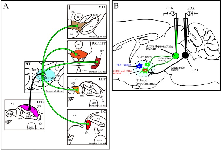
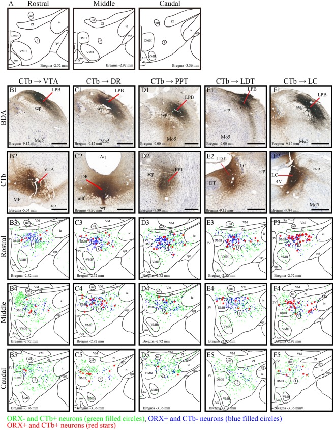
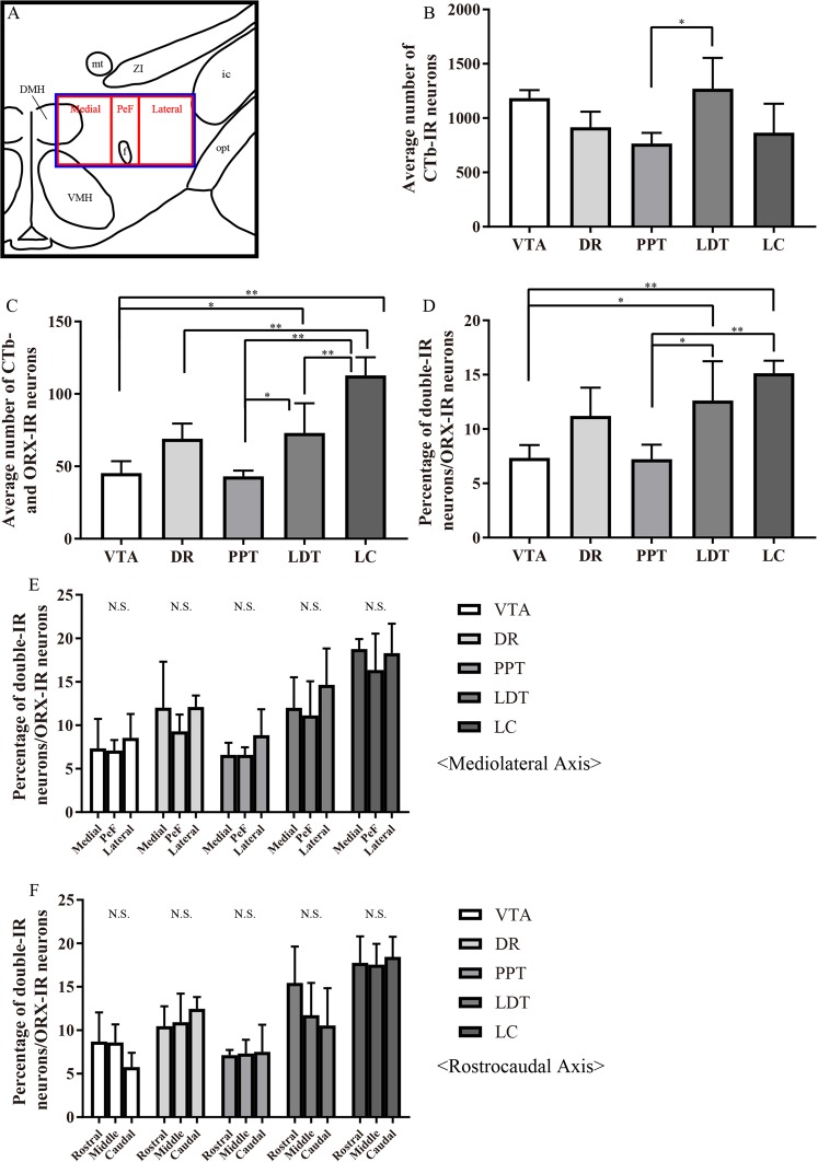
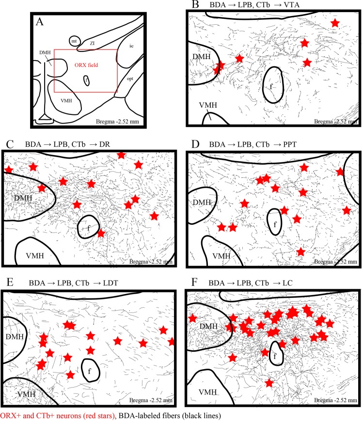
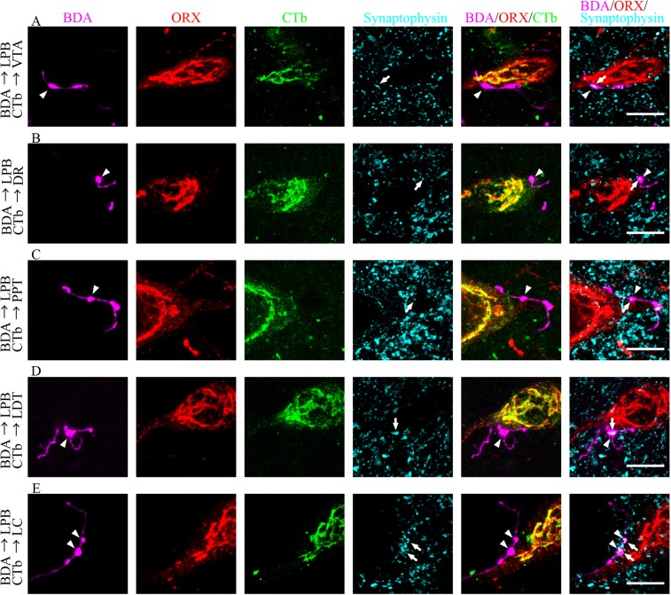
Orexin (ORX) neurons in the hypothalamus send their axons to arousal-promoting areas. We have previously shown that glutamatergic neurons in the lateral parabrachial nucleus (LPB) innervate ORX neurons. In this study, we examined potential pathways from the LPB to ORX neurons projecting to arousal-promoting areas in the brainstem by a combination of tract-tracing techniques in male Wistar rats. We injected the anterograde tracer biotinylated dextranamine (BDA) into the LPB and the retrograde tracer cholera toxin B subunit (CTb) into the ventral tegmental area, dorsal raphe nucleus, pedunculopontine tegmental nucleus, laterodorsal tegmental area, or locus coeruleus (LC). We then analyzed the BDA-labeled fibers and ORX-immunoreactive neurons in the hypothalamus. We found that double-labeled ORX and CTb neurons were the most abundant after CTb was injected into the LC. We also observed prominently overlapping distribution of BDA-labeled fibers, arising from neurons located in the lateral-most part of the dorsomedial nucleus and adjacent dorsal perifornical area. In these areas, we confirmed by confocal microscopy that BDA-labeled synaptophysin-immunoreactive axon terminals were in contiguity with cell bodies and dendrites of CTb-labeled ORX-immunoreactive neurons. These results suggest that the LPB innervates arousal-promoting areas via ORX neurons and is likely to promote arousal responses to stimuli.


Conflict of interest statement

The authors declare no competing interests.

Figures

Figure 1
Figure 1
Schematic diagram of this research. (A) Diagram of axonal projections from LPB to brainstem arousal areas via ORX neurons which were newly identified in this study. (B) Schematic drawings of combined anterograde (black) and retrograde (green) tract-tracing method. Immunohistochemistry was performed on sections of the tuberal hypothalamus (dotted line). CTb negative and ORX positive neurons (ORX+) are represented as blue neurons, CTb positive ORX negative (CTb+) cells are as green neurons and CTb positive ORX positive neurons (ORX+ and CTb+) are as red neurons. LPB; lateral parabrachial nucleus, ORX; orexin, CTb; cholera toxin subunit B, BDA; biotinylated dextranamine, VTA; ventral tegmental area, PPT; pedunculopontine tegmental nucleus, LDT; laterodorsal tegmental area, LC; locus coeruleus, DR; dorsal raphe nucleus.
Figure 2
Figure 2
Distribution of ORX-immunoreactive (IR) neurons projecting to the brainstem arousal areas. (A) Schematic drawings of three sections which represent the approximate posterior levels to bregma −2.54 mm (rostral), −2.92 mm (middle) and −3.36 mm (Caudal). (B) Photomicrographs showing the injection sites of BDA into the LPB (B1-F1) and CTb into the VTA (B2), DR (C2), PPT (D2), LDT (E2) and LC (F2). (B3- F5) Line drawings showing the distribution of CTb-IR neurons (green filled circles), ORX-IR neurons (blue filled circles), and ORX and CT- double-IR neurons (red stars) in the three regions of hypothalamus. The distance (mm) behind the bregma is noted at the bottom. Scale bars, 500 μm. CTb; cholera toxin b subunit; ORX: orexin; IR: immunoreactive; VTA: ventral tegmental area; DR: dorsal raphe; PPT: pedunculopontine nucleus; LDT: laterodorsal tegmental nucleus; LC: locus ceruleus; BDA: biotinylated dextranamine.
Figure 3
Figure 3
Abundant innervation of hypothalamic orexin neurons to LC. (A) Schematic drawing showing arbitrarily divided three regions of the hypothalamus. Blue square shows ORX field and this square is divided to three regions, medial, PeF, and lateral. (BD) Quantification of the average number of CTb-labeled neurons (B), ORX-IR neurons and CTb-labeled neurons (C), and the percentage of ORX and CTb double-IR neurons in ORX-IR neurons (D) in the ORX field. *P < 0.05, **P < 0.01, (n = 4, 6 sections from each rat. one-way ANOVA followed by Tukey–Kramer test) (E and F) Quantification of the percentage of ORX and CTb double-IR neurons in ORX-IR neurons after an injection of CTb into the VTA, DR, PPT, LDT or LC. Cells were quantified in three rectangles, medial, PeF and lateral regions across mediolateral axis, and rostral, middle and caudal rectangles in the ORX field of the hypothalamus across the rostrocaudal axis and compared among samples. N.S.; not significantly different. PeF: perifornical area; CTb; cholera toxin b subunit; ORX: orexin; IR: immunoreactive; VTA: ventral tegmental area; DR: dorsal raphe; PPT: pedunculopontine nucleus; LDT: laterodorsal tegmental nucleus; LC: locus coeruleus.
Figure 4
Figure 4
Distribution of LPB axons and ORX and CTb double-IR neurons in the ORX field. (A) Schematic drawing shows the ORX field. The distance (mm) behind the bregma is noted at the bottom right. (BF) Line drawings showing the distribution of BDA-labeled axons (black line), and ORX and CTb double-IR neurons (red stars) in the hypothalamus. The distance (mm) behind the bregma is noted at the bottom right. LPB: lateral parabrachial nuclei; ORX: orexin; CTb: cholera toxin b subunit; BDA: biotinylated dextranamine.
Figure 5
Figure 5
Confocal microscope images of quadruple fluorescence staining. Images show the appearance of CTb-labeled neurons (green) immunoreactive for ORX (red) and Synaptophysin (cyan) after injection of BDA (magenta) into the LPB and CTb into the VTA (A) DR (B) PPT (C) LDT (D) or LC (E) Two types of merged images, BDA/ORX/CTb and BDA/ORX/synaptophysin, were obtained from the same area. The arrowheads indicate BDA-labeled axons (magenta), which contact to ORX (red) and CTb (green) double-IR neurons. The arrows indicate synaptophysin (cyan) in BDA-labeled axons (magenta), which contact to ORX (red)-IR neurons. Scale bars, 10 μm. LPB: lateral parabrachial nuclei; ORX: orexin; VTA: ventral tegmental area; IR: immunoreactive; CTb: cholera toxin B subunit; BDA: biotinylated dextranamine; PPT: pedunculopontine tegmental nucleus; VTA: ventral tegmental area; DR: dorsal raphe; LC: locus coeruleus.

References

    1. Sakurai T, et al. Orexins and orexin receptors: a family of hypothalamic neuropeptides and G protein-coupled receptors that regulate feeding behavior. Cell. 1998;92:573–585. doi: 10.1016/S0092-8674(00)80949-6. - DOI - PubMed
    1. Mieda M. The roles of orexins in sleep/wake regulation. Neurosci. Res. 2017;118:56–65. doi: 10.1016/j.neures.2017.03.015. - DOI - PubMed
    1. de Lecea L, et al. The hypocretins: hypothalamus-specific peptides with neuroexcitatory activity. Proc. Natl. Acad. Sci. USA. 1998;95:322–327. doi: 10.1073/pnas.95.1.322. - DOI - PMC - PubMed
    1. Nambu T, et al. Distribution of orexin neurons in the adult rat brain. Brain Res. 1999;827:243–260. doi: 10.1016/S0006-8993(99)01336-0. - DOI - PubMed
    1. Peyron C, et al. Neurons containing hypocretin (orexin) project to multiple neuronal systems. J. Neurosci. 1998;18:9996–10015. doi: 10.1523/JNEUROSCI.18-23-09996.1998. - DOI - PMC - PubMed