

Proton magnetic resonance spectroscopy (1H-MRS) of the brain in patients with tick-borne encephalitis
- PMID: 30808997
- PMCID: PMC6391410
- DOI: 10.1038/s41598-019-39352-6
Proton magnetic resonance spectroscopy (1H-MRS) of the brain in patients with tick-borne encephalitis
Abstract
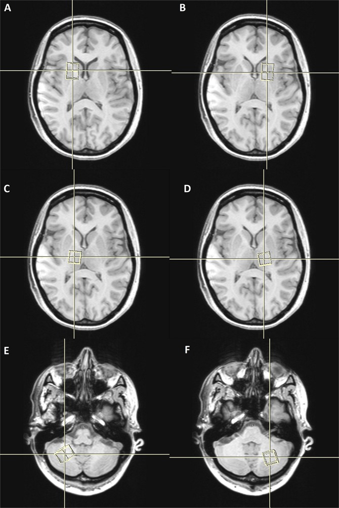
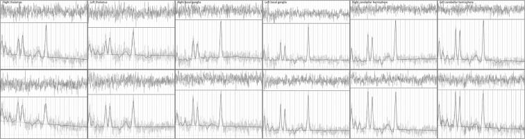
Tick-borne encephalitis (TBE) is a disease caused by a tick-borne encephalitis virus (TBEV) belonging to the Flaviviridae family. The aforementioned virus is transmitted by the bite of infected ticks. In the recent years, TBEV has become a serious public health problem with a steady increase in its incidence, mainly due to the climate changes and spreading the infected ticks into new territories. The standard protocol of TBE diagnosis involves the serological laboratory test with a minor role of imaging techniques such as magnetic resonance imaging. Long-term complications affecting patients daily activities are reported in about 40-50% of the cases. However, no changes are revealed in the laboratory tests or the imaging examination. The development of new imaging techniques such as proton magnetic resonance spectroscopy (1H-MRS) can broaden the knowledge about TBE, contributing to its prevention. The aim of this study was to assess the usefulness of 1H-MRS of the brain in patients with TBE. Compared to controls, a statistically significant decrease in the N-acetylaspartate /creatine ratio was found bilaterally in the right and left thalamus as well as a statistically significant increase in the choline/creatine ratio in the right and left thalamus.
Conflict of interest statement
The authors declare no competing interests.
Figures
Similar articles
-
COMPARISON BETWEEN PROTON MAGNETIC RESONANCE SPECTROSCOPY FINDINGS IN DOGS WITH TICK-BORNE ENCEPHALITIS AND CLINICALLY NORMAL DOGS.Vet Radiol Ultrasound. 2017 Jan;58(1):53-61. doi: 10.1111/vru.12427. Epub 2016 Oct 7. Vet Radiol Ultrasound. 2017. PMID: 27714889
-
Cerebral glucose hypometabolism in Tick-Borne Encephalitis, a pilot study in 10 Patients.Int J Infect Dis. 2016 Oct;51:73-77. doi: 10.1016/j.ijid.2016.06.022. Epub 2016 Jul 11. Int J Infect Dis. 2016. PMID: 27418580
-
Patients with breakthrough tick-borne encephalitis suffer a more severe clinical course and display extensive magnetic resonance imaging changes.Eur J Neurol. 2020 Jul;27(7):1201-1209. doi: 10.1111/ene.14276. Epub 2020 May 19. Eur J Neurol. 2020. PMID: 32324925 Free PMC article.
-
EAN consensus review on prevention, diagnosis and management of tick-borne encephalitis.Eur J Neurol. 2017 Oct;24(10):1214-e61. doi: 10.1111/ene.13356. Epub 2017 Aug 1. Eur J Neurol. 2017. PMID: 28762591 Review.
-
Tick-borne encephalitis in Europe and Russia: Review of pathogenesis, clinical features, therapy, and vaccines.Antiviral Res. 2019 Apr;164:23-51. doi: 10.1016/j.antiviral.2019.01.014. Epub 2019 Jan 31. Antiviral Res. 2019. PMID: 30710567 Review.
Cited by
-
Meta-analysis and Open-source Database for In Vivo Brain Magnetic Resonance Spectroscopy in Health and Disease.bioRxiv [Preprint]. 2023 Jun 15:2023.02.10.528046. doi: 10.1101/2023.02.10.528046. bioRxiv. 2023. Update in: Anal Biochem. 2023 Sep 1;676:115227. doi: 10.1016/j.ab.2023.115227. PMID: 37205343 Free PMC article. Updated. Preprint.
-
Meta-analysis and open-source database for in vivo brain Magnetic Resonance spectroscopy in health and disease.Anal Biochem. 2023 Sep 1;676:115227. doi: 10.1016/j.ab.2023.115227. Epub 2023 Jul 7. Anal Biochem. 2023. PMID: 37423487 Free PMC article.
References
-
- World Health Organization. Vaccines against tick-borne encephalitis: WHO position paper. Wkly Epidemiol Rec. 86, 241–56 (2011). - PubMed
Publication types
MeSH terms
Substances
LinkOut - more resources
Full Text Sources
