Clinical Trial
. 2019 Jul;27(7):1054-1060.
doi: 10.1038/s41431-019-0357-x. Epub 2019 Feb 26.

SMAD4 rare variants in individuals and families with thoracic aortic aneurysms and dissections

Affiliations
Clinical Trial

SMAD4 rare variants in individuals and families with thoracic aortic aneurysms and dissections

Xue-Yan Duan et al. Eur J Hum Genet. 2019 Jul.

Abstract

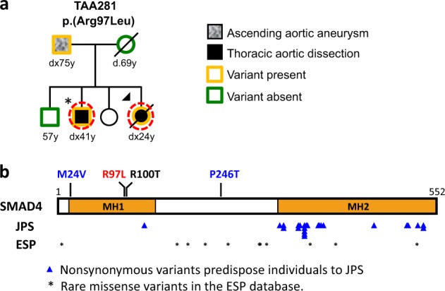
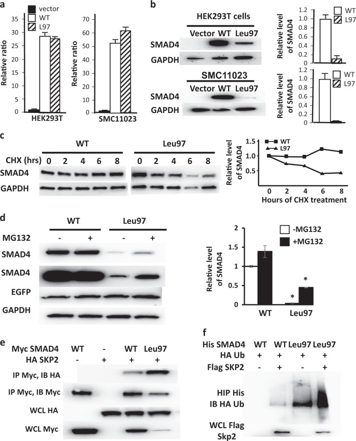
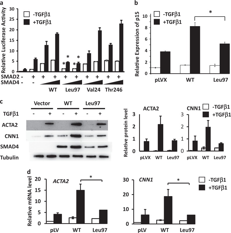
SMAD4 pathogenic variants cause juvenile polyposis (JPS) and hereditary hemorrhagic telangiectasia (HHT), and 40% of affected individuals also have thoracic aortic disease. At the same time, SMAD4 pathogenic variants have not been reported in thoracic aortic disease families without JPS-HHT. A SMAD4 heterozygous variant, c.290G>T, p.(Arg97Leu), not present in population databases and predicted to be damaging to protein function, was identified in a family with thoracic aortic disease and no evidence of HHT or JPS. Cellular studies revealed that the SMAD4 p.(Arg97Leu) alteration increased SMAD4 ubiquitination and 26S proteasome-mediated protein degradation. Smooth muscle cells (SMCs) infected with lentivirus expressing the SMAD4 p.(Arg97Leu) variant demonstrated reduced contractile protein gene expression when compared to that of wild-type SMAD4. In addition, two rare variants were identified in individuals with early age of onset of thoracic aortic dissection. These results suggest that SMAD4 rare missense variants can lead to thoracic aortic disease in individuals who do not have JPS or HHT.


Conflict of interest statement

The authors declare that they have no conflict of interest.

Figures

Fig. 1
Fig. 1
SMAD4 rare variants identified in a family with heritable thoracic aortic disease. a Pedigree of TAA281 with SMAD4 p.(Arg97Leu) variant. The legend indicates the disease status and genotypes of the family members. Arrow points to the proband. Asterisk indicates the presence of a bicuspid aortic valve. The age at diagnosis of aortic aneurysm or dissection (‘‘dx’’) and age at death (‘‘d’’) are shown in years. A dashed circle around a symbol indicates individuals whose DNA was used for exome sequencing. b Schematic representation of the SMAD4 domains and variants. The SMAD4 rare missense variant identified in TAA281 is shown in red, the somatic variant identified in pancreatic cancer in black, and missense variants identified in individuals with early dissections are in blue above the protein diagram. The blue triangles indicate the location of SMAD4 missense variants identified in patients with JPS or JPS–HHT. Asterisks indicate variants identified in the NHLBI ESP database
Fig. 2
Fig. 2
SMAD4 Leu97 reduces protein stability through increased binding to SKP2. a Lentiviruses of control vector and Flag-tagged WT and Leu97 SMAD4 were infected in HEK293T and SMC11023 followed by real-time qPCR with GAPDH as internal control. SMAD4 WT and Leu97 have similar expression of mRNA. b In these same cells, the protein level of SMAD4 Leu97 was significantly lower than that of SMAD4 WT. The average protein levels over triplicate experiments are shown on the graph. c SMAD4 Leu97 variant protein is unstable after cycloheximide treatment. HEK293T cells expressing SMAD4 WT and Leu97 variant were treated with cycloheximide for up to 8 h followed by immunoblot analyses with anti-Flag antibodies. The levels of SMAD4 and GAPDH protein were quantified using immunoblot assay. d MG132 partially rescued the degradation of SMAD4 Leu97 variant. HEK293T cells expressing SMAD4 WT and Leu97 variant were treated with MG132, for 8 h. A representative experiment is shown on the left and quantification of three individual experiments are shown at the right. e Leu97 variant increased the ability of SMAD4 binding to HA-SKP2. HEK293T cells were co-transfected with Myc-SMAD4 and HA-SKP2 followed by immunoprecipitation with Myc beads. The SMAD4-bound SKP2 was detected with anti-HA antibodies. f Ubiquitination of SMAD4 Leu97 is increased compared to WT protein. HEK293T cells were co-transfected with His-SMAD4, Flag-SKP2, and HA-ubiquitin followed by immunoprecipitation with Ni-NTA agarose beads under denatured conditions. The ubiquitination of Smad4 was detected with anti-HA antibodies. Three individual experiments were performed and one representative result is shown. Asterisks indicate a p value <0.05
Fig. 3
Fig. 3
SMAD4 Leu97 variant reduces TGFβ signaling. a The effect of SMAD4 rare variants on the TGFβ was assessed in COS7 cells signaling. The SMAD4 activity of Leu97 alteration to induce 3TP luciferase activity was reduced compared to WT. COS7 cells were co-transfected with 3TP-lux plasmid and plasmids expressing WT or rare variants in SMAD4, followed by TGFβ1 treatment for 24 h. The 3TP luciferase activities were measured with renilla luciferase as internal control. The enhancement of luciferase activity in response to TGFβ1 treatment in SMAD4 Leu97 was significantly lower than that of SMAD4 WT, Val24, Thr246. b Immortalized SMCs (SMC11023) expressing control vector, SMAD4 WT or SMAD4 Leu97 were treated with TGFβ1 for 4 h and the expression of p15INK4b was measured by real-time qPCR and with GAPDH as internal control. c, d SMAD4 Leu97 variant reduced TGFβ1-induced smooth muscle specific gene expression and protein levels. SMC11023 cells infected with control vector, Flag-tagged SMAD4 WT or Leu97 variant were treated with TGFβ1 for 48 h followed by Western blot (c) and real-time qPCR (d). Asterisks indicate a p value <0.05 when compared to the SMAD4 WT

References

    1. Howard DP, Banerjee A, Fairhead JF, Perkins J, Silver LE, Rothwell PM. Population-based study of incidence and outcome of acute aortic dissection and premorbid risk factor control: 10-year results from the Oxford Vascular Study. Circulation. 2013;127:2031–7. doi: 10.1161/CIRCULATIONAHA.112.000483. - DOI - PMC - PubMed
    1. Prakash SK, Haden-Pinneri K, Milewicz DM. Susceptibility to acute thoracic aortic dissections in patients dying outside the hospital: an autopsy study. Am Heart J. 2011;162:474–9. doi: 10.1016/j.ahj.2011.06.020. - DOI - PubMed
    1. Hiratzka LF, Bakris GL, Beckman JA, et al. 2010 ACCF/AHA/AATS/ACR/ASA/SCA/SCAI/SIR/STS/SVM guidelines for the diagnosis and management of patients with Thoracic Aortic Disease: a report of the American College of Cardiology Foundation/American Heart Association Task Force on Practice Guidelines, American Association for Thoracic Surgery, American College of Radiology, American Stroke Association, Society of Cardiovascular Anesthesiologists, Society for Cardiovascular Angiography and Interventions, Society of Interventional Radiology, Society of Thoracic Surgeons, and Society for Vascular Medicine. Circulation. 2010;121:e266–369. - PubMed
    1. Mizuguchi T, Collod-Beroud G, Akiyama T, et al. Heterozygous TGFBR2 mutations in Marfan syndrome. Nat Genet. 2004;36:855–60. doi: 10.1038/ng1392. - DOI - PMC - PubMed
    1. Loeys BL, Schwarze U, Holm T, et al. Aneurysm syndromes caused by mutations in the TGF-beta receptor. N Engl J Med. 2006;355:788–98. doi: 10.1056/NEJMoa055695. - DOI - PubMed

Publication types