Guideline
. 2019 Feb 21;25(7):744-776.
doi: 10.3748/wjg.v25.i7.744.

Consensus on the digestive endoscopic tunnel technique

Affiliations
Guideline

Consensus on the digestive endoscopic tunnel technique

Ning-Li Chai et al. World J Gastroenterol. .

Abstract

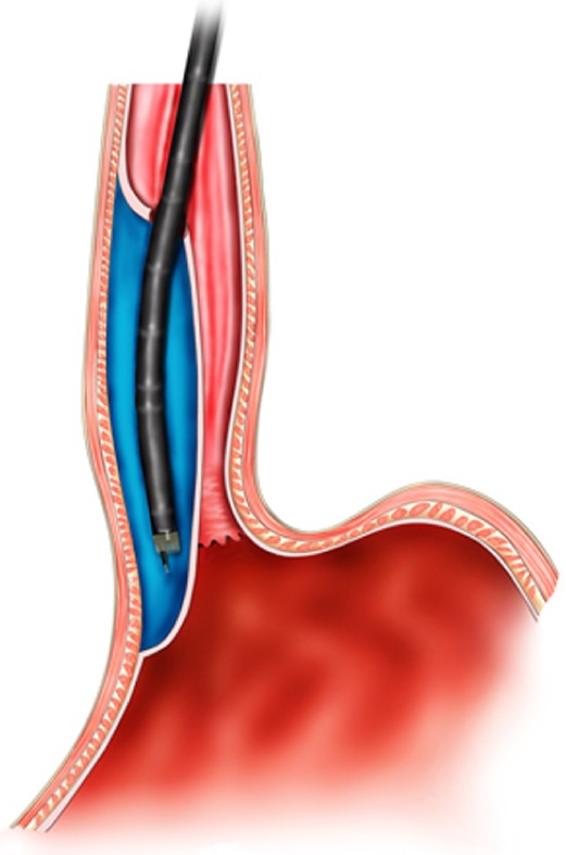
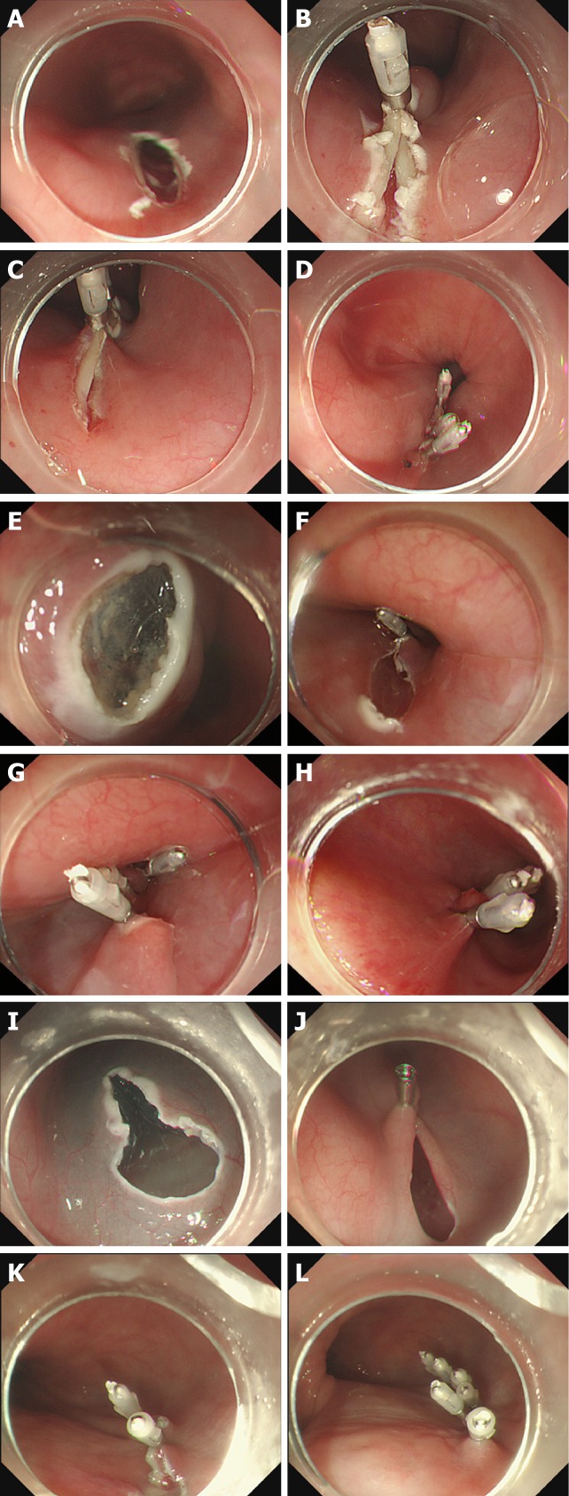
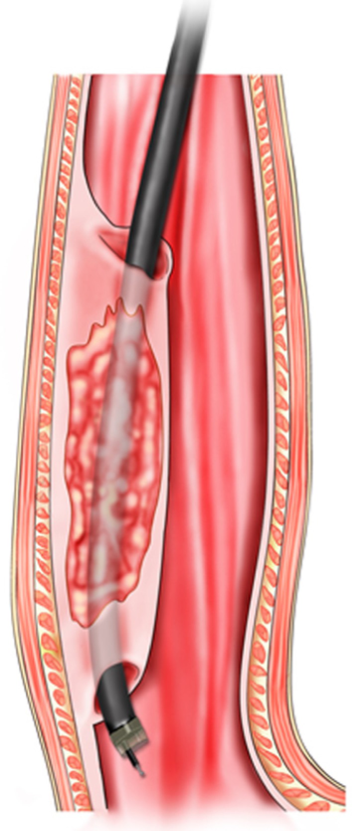
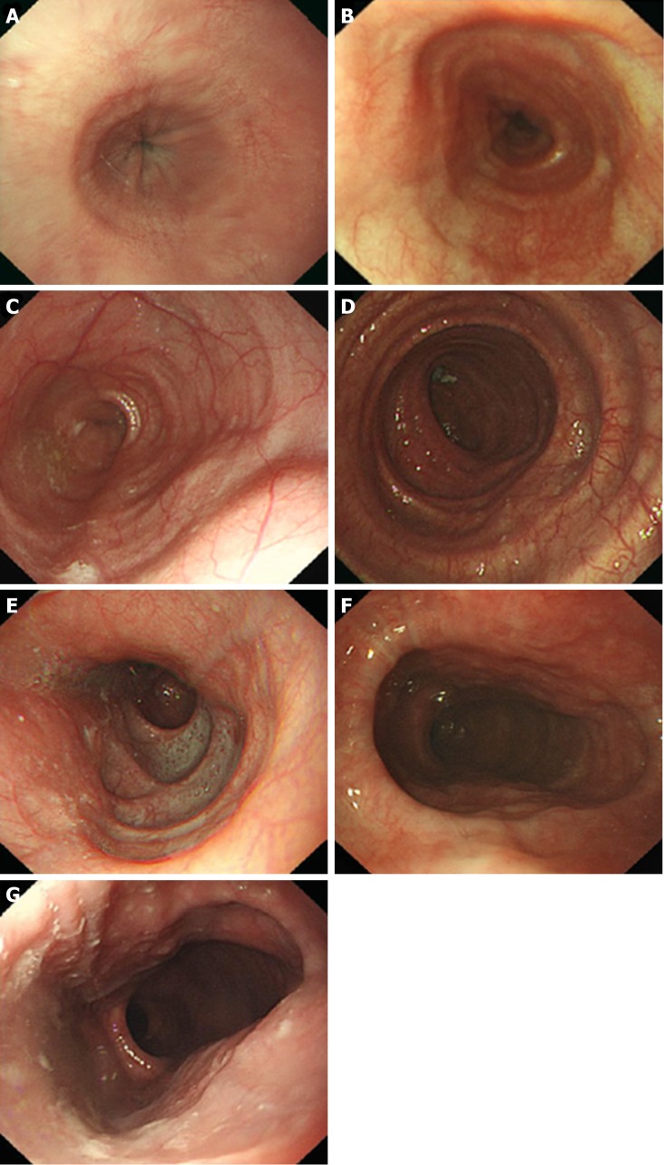
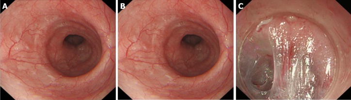
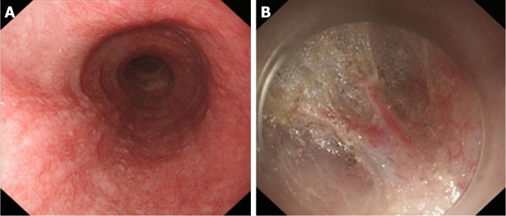
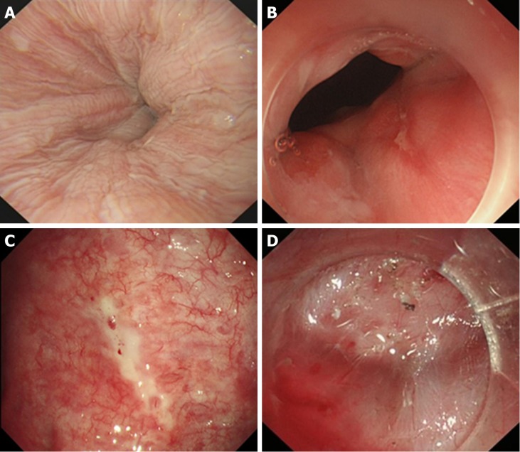
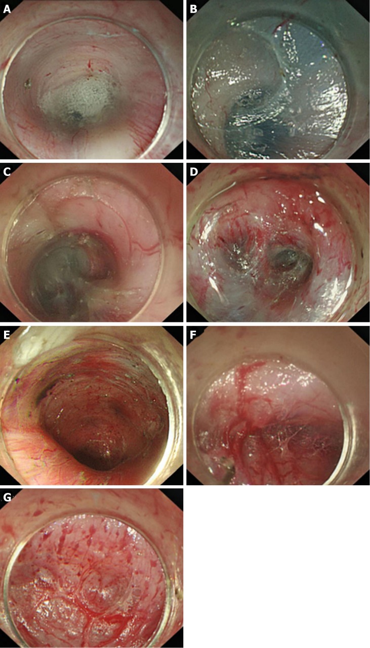
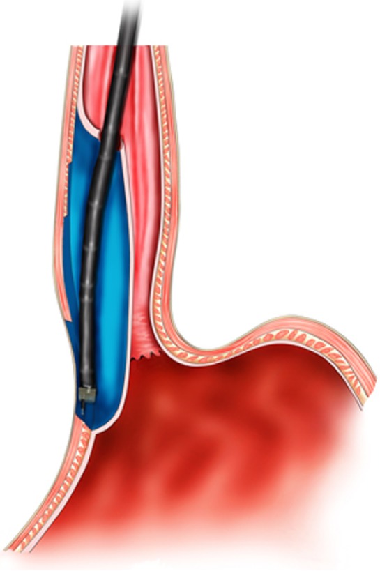
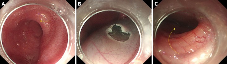
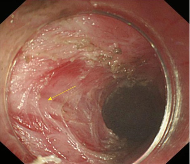
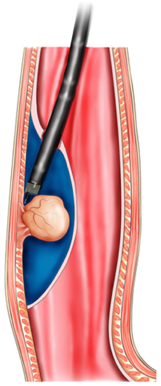
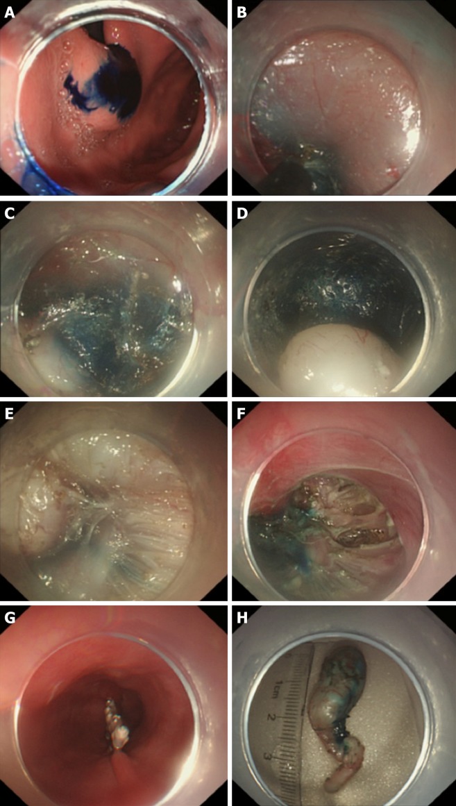
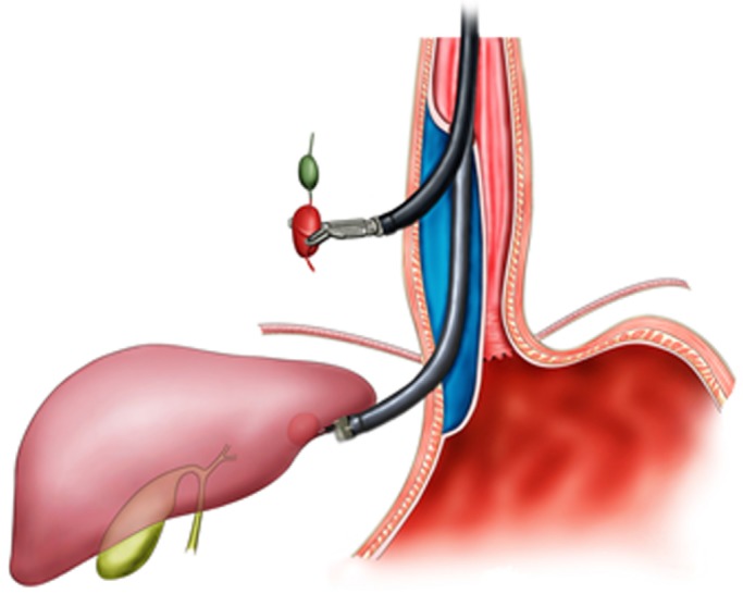
With the digestive endoscopic tunnel technique (DETT), many diseases that previously would have been treated by surgery are now endoscopically curable by establishing a submucosal tunnel between the mucosa and muscularis propria (MP). Through the tunnel, endoscopic diagnosis or treatment is performed for lesions in the mucosa, in the MP, and even outside the gastrointestinal (GI) tract. At present, the tunnel technique application range covers the following: (1) Treatment of lesions originating from the mucosal layer, e.g., endoscopic submucosal tunnel dissection for oesophageal large or circular early-stage cancer or precancerosis; (2) treatment of lesions from the MP layer, per-oral endoscopic myotomy, submucosal tunnelling endoscopic resection, etc.; and (3) diagnosis and treatment of lesions outside the GI tract, such as resection of lymph nodes and benign tumour excision in the mediastinum or abdominal cavity. With the increasing number of DETTs performed worldwide, endoscopic tunnel therapeutics, which is based on DETT, has been gradually developed and optimized. However, there is not yet an expert consensus on DETT to regulate its indications, contraindications, surgical procedure, and postoperative treatment. The International DETT Alliance signed up this consensus to standardize the procedures of DETT. In this consensus, we describe the definition, mechanism, and significance of DETT, prevention of infection and concepts of DETT-associated complications, methods to establish a submucosal tunnel, and application of DETT for lesions in the mucosa, in the MP and outside the GI tract (indications and contraindications, procedures, pre- and postoperative treatments, effectiveness, complications and treatments, and a comparison between DETT and other operations).

Keywords: Digestive endoscopic tunnel technique; Endoscopic submucosal tunnel dissection; Gastrointestinal tract; Per-oral endoscopic myotomy; Submucosal tunnelling endoscopic resection.


Conflict of interest statement

Conflict-of-interest statement: All authors have no financial relationships to disclose.

Figures

Figure 1
Figure 1
Mechanism of digestive endoscopic tunnel technique, demonstrating a tunnel that is created between the mucosal and muscularis propria layers.
Figure 2
Figure 2
Three methods of tunnel incision and closure. A and B: Longitudinal incision; C and D: Longitudinal incision closed with titanium clips; E: Transverse incision; F: “Anchoring” of a titanium clip in the middle of the transverse incision; G and H: Longitudinal closure using titanium clips successively; I: Inverted T incision; J-L: Longitudinal closure using titanium clips successively.
Figure 3
Figure 3
Schema chart of endoscopic submucosal tunnel dissection, demonstrating a tunnel that is created to resect mucosal lesions.
Figure 4
Figure 4
Simulated diagram of endoscopic observations in Ling IIb and Ling IIc. A: Ling IIb. The arrows indicate 1/3 of the oesophageal cavity, and the semi-annular structure’s midpoint remains within this range; B: Ling IIc. The arrows indicate 1/3 of the oesophageal cavity, and the crescent-like structure's midpoint goes beyond this range.
Figure 5
Figure 5
Endoscopic images of the Ling classification of achalasia cardia. A: Ling I; B: Ling IIa; C: Ling IIb; D: Ling IIc, E: Ling III1; F: Ling IIIr; G: Ling IIIlr.
Figure 6
Figure 6
Correlation between grade A or grade B mucosal inflammation and mild oesophageal submucosal adhesion. A: Grade A mucosal inflammation; B: Grade B mucosal inflammation; C: Mild oesophageal submucosal adhesion: The fibre filaments are distributed in bundles.
Figure 7
Figure 7
Correlation between Grade C mucosal inflammation and moderate oesophageal submucosal adhesion. A: Grade C mucosal inflammation; B: Moderate oesophageal submucosal adhesion. The fibres are arranged in disorder, with fusion and decreased transparency.
Figure 8
Figure 8
Correlation between Grade D, Grade E, or Grade F mucosal inflammation and severe oesophageal submucosal adhesion. A: Grade D mucosal inflammation; B: Grade E mucosal inflammation; C: Grade F mucosal inflammation; D: Severe oesophageal submucosal adhesion. The submucosa and muscularis propria are completely adherent.
Figure 9
Figure 9
Anatomical landmark in the tunnel from the lower oesophagus to the cardia. A: Grid-like blood vessels in the cardia; B: Crescent-like structure visible at the proximal cardia; C: Ampulla-like structure appearing after entering the crescent-like structure; D: Branching vessels with bulky vascular roots in the ampulla-like structure; E: Tunnel below the cardia, showing a steep downward form; F: Stubby and multi-branched vessels below the cardia; G: Beadlike vessels below the cardia.
Figure 10
Figure 10
Schema chart of per-oral endoscopic myotomy, demonstrating a tunnel that is created to incise the muscularis propria.
Figure 11
Figure 11
Endoscope crosses the “ridge” via short-tunnel per-oral endoscopic myotomy. A: Type Ling IIc oesophagus. The arrow indicates a “ridge” structure formed by the crescent-like structure; B: The short-tunnel entry incision established on a relatively flat oesophageal wall at the oral side of the “ridge”; C: It is easy to cross the “ridge” within the tunnel.
Figure 12
Figure 12
Inner circular muscle myotomy. The arrow shows the well-retained longitudinal muscle.
Figure 13
Figure 13
Full-thickness myotomy. The arrow shows the tunica adventitia of the oesophagus.
Figure 14
Figure 14
Glasses-style myotomy. The arrow shows the muscles remaining at the cardia.
Figure 15
Figure 15
Circular muscle myotomy + balloon plasty. A: The width of the incision should be 1/3 of the circumferential oesophagus; B: Balloon-dilation in the oesophagus; C: The width of the incision after expansion should be 2/3 of the circumferential oesophagus.
Figure 16
Figure 16
Progressive full-thickness myotomy. The yellow arrow shows the incision into the inner circular muscles from superficial to deep; the blue arrow shows the full incision into the muscularis propria.
Figure 17
Figure 17
Schema chart of per-oral endoscopic myotomy, demonstrating a tunnel that is created to resect the lesion from the muscularis propria.
Figure 18
Figure 18
Key steps of submucosal tunnelling endoscopic resection for a cardial submucosal tumour. A: Injection of methylene blue into the lesion incision site for marking and positioning; B: Establishment of a tunnel; C: Finding of the marking and positioning with methylene blue in the tunnel; D: Exposure of the lesion; E: Resection of the lesion; F: Wound following the resection of the tumour; G: Closure of the mucosal incision; H: The resected specimen.
Figure 19
Figure 19
Schema chart of digestive endoscopic tunnel technique on the external digestive tract wall, demonstrating a tunnel that is created to resect a node outside the digestive tract.

Similar articles

Cited by

References

    1. Linghu E. Endoscopic resection for gastrointestinal pre-cancerous lesion and early cancer. Electronic Image Press of the Chinese Medical Association. 2009.
    1. Linghu E. Therapeutics of digestive endoscopic tunnel technique. Berlin: Springer; 2014. pp. 1–3.
    1. Linghu E. The establishment and prospect of endoscopic tunnel technique. Zhonghua Qiangjing Waike Zazhi (Electronic Edition) 2011;4:1–2.
    1. Linghu E. A view of the principle and base of endoscopic technique innovation from the development of endoscopic submucosa dissection and peroral endoscopic myotomy. Zhonghua Xiaohua Neijing Zazhi. 2011;28:603–604.
    1. Werner YB, Rösch T. POEM and Submucosal Tunneling. Curr Treat Options Gastroenterol. 2016;14:163–177. - PubMed

Publication types

MeSH terms