. 2019 Feb 11:12:1137-1146.
doi: 10.2147/OTT.S190274. eCollection 2019.

Epstein-Barr virus-encoded LMP1 regulated Pim1 kinase expression promotes nasopharyngeal carcinoma cells proliferation

Affiliations

Epstein-Barr virus-encoded LMP1 regulated Pim1 kinase expression promotes nasopharyngeal carcinoma cells proliferation

Ran-Ran Ding et al. Onco Targets Ther. .

Abstract

Background: Epstein-Barr virus-encoded LMP1 plays a critical role in the carcinogenesis of nasopharyngeal carcinoma (NPC), but the mechanism remains elusive. We aimed to analyze the expression and clinical pathological significance of provirus integration site for Moloney murine leukemia virus 1 (Pim1) in clinical NPC, and to elucidate the effect of LMP1 on Pim1 expression and its mechanism.

Methods: Immunohistochemical staining was used to detect the expression of Pim1 in clinical NPC tissues and control nasopharyngeal chronic inflammation (NPI) tissues, and the correlation between Pim1 and clinical parameters of NPC patients was analyzed. The LMP1 stable expression cell line CNE1-LMP1-OV was constructed through infecting the well-differentiated nasopharyngeal carcinoma cells CNE1 with LMP1 overexpressing lentivirus. Then the in vivo experiments were conducted.

Results: Among 89 NPC patients, 48 cases (53.93%) were positive for Pim1, while only one case was Pim1 positive in 15 NPI controls (6.67%). Pim1 expression was not correlated with gender, age, smoking status and clinical classification of NPC patients, but positively correlated with T, N and M classification. CNE1-LMP1-OV cell line was successfully established, which displayed a higher cell proliferation ability and Pim1 expression. NF-κB inhibitor PDTC, PKC inhibitor GF109203X and STAT3 inhibitor Stattic significantly attenuated LMP1-induced Pim1 expression, and while AP-1 inhibitor SR11302 showed no inhibitory effect. Interestingly, Pim1 inhibitor quercetagetin significantly inhibited the proliferation of CNE1-LMP1-OV cells.

Conclusion: LMP1 mediates Pim1 expression through NF-κB, PKC and STAT3 signaling, which promotes the proliferation of NPC cells and participate in the clinical progression of NPC.

Keywords: LMP1; Pim1; cell proliferation; nasopharyngeal carcinoma.


Conflict of interest statement

Disclosure The authors report no conflicts of interest in this work.

Figures

Figure 1
Figure 1
Pim1 was upregulated in NPC clinical specimens. Notes: (A) Pim1 immunohistochemistry. Representative images of Pim1 expression in clinical specimens. PBS substituted for primary antibody was served as blank control, nuclei were counterstained with hematoxylin. Bars, 100 µm. (B) Representative immunofluorescence images of Pim1 in NPC and NPI clinical specimens. Nuclei were counterstained with DAPI, and antigens for Pim1 were colorized with rhodamine-coupled IgGs. Bars, 50 µm. Abbreviations: DAPI, 4′,6-diamidino-2-phenylindole; NPC, nasopharyngeal carcinoma; NPI, nasopharyngeal chronic inflammation.
Figure 2
Figure 2
Establishment of LMP1 overexpressing cell line. Notes: (A) Efficiency of lentiviral infection in CNE1 cells. LMP1 overexpressing lentiviral particles and control lentiviral particles infected the CNE1 cells at MOI of 20, 3 days post infection, and the eGFP signal was observed under a fluorescence microscope. Bars, 200 µm. (B) Detection of LMP1 in lentivirus-infected CNE1 cells by immunofluorescence staining. Nuclei were counterstained with DAPI, and antigens for LMP1 were colorized with FITC-coupled IgGs. Bars, 100 µm. (C) Immunoblotting of LMP1 protein in established cell lines. (D) Semiquantitative results of LMP1. The experiment was repeated three times (n=3). ***P<0.001. Abbreviation: DAPI, 4′,6-diamidino-2-phenylindole.
Figure 3
Figure 3
LMP1 induces PCNA expression in NPC cells. Notes: (A) Detection of PCNA in lentivirus-infected CNE1 cells by immunofluorescence staining. Nuclei were counterstained with DAPI, and antigens for PCNA were colorized with TRITC-coupled IgGs. Bars, 50 µm. (B) Immunoblotting of PCNA in established cell line. The experiment was repeated three times (n=3) and the representative bands are shown. ***P<0.001. Abbreviations: DAPI, 4′,6-diamidino-2-phenylindole; PCNA, proliferating cell nuclear antigen; TRITC, tetramethylrhodamine isothiocyanate.
Figure 4
Figure 4
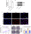
LMP1-induced Pim1 expression promotes NPC cell proliferation. Notes: (A) Enhanced Pim1 mRNA levels in LMP1 stably expressing cells. ***P<0.001. The experiment was repeated three times (n=3). (B) LMP1 mainly upregulates the 33 kDa Pim1 protein but not the 44 kDa protein in NPC cells. The experiment was repeated three times (n=3) and the representative bands are shown. (C) Expression and cellular location of Pim1 protein in NPC cells. Antigens for Pim1 were colorized with TRITC-coupled IgGs. Bars, 75 µm. (D) Cell viability of LMP1-overexpressing cells and control cells treated with Pim1 inhibitor quercetagetin, assessed by CCK-8 assay. *P<0.05. Each group was repeated in six wells (n=6). (E) Clone formation efficiency of quercetagetin-treated cells. *P<0.05. Each group was repeated in three wells (n=3). Abbreviations: CCK-8, cell counting kit-8; DAPI, 4′,6-diamidino-2-phenylindole; DMSO, dimethyl sulfoxide; NS, no significance; TRITC, tetramethylrhodamine isothiocyanate.
Figure 5
Figure 5
Signaling involved in LMP1-induced Pim1 expression in NPC cells. Notes: (A) Pim1 mRNA levels were detected in LMP1 downstream signaling inhibitors including PDTC, GF109203X, Stattic and SR11302. *P<0.05, ***P<0.001; PDTC, NF-κB inhibitor; GF109203X, PKC inhibitor; Stattic, STAT3 inhibitor; SR11302, AP-1 inhibitor. The experiment was repeated three times (n=3). (B) Pim1 protein levels were detected in LMP1 downstream signaling inhibitors including PDTC, GF109203X, Stattic and SR11302. *P<0.05, ***P<0.001. The experiment was repeated three times (n=3) and the representative bands are shown. Abbreviations: DMSO, dimethyl sulfoxide; PDTC, pyrrolidine dithiocarbamate.
Figure 6
Figure 6
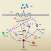
Schematic illustration of the mechanism of LMP1/Pim1 signaling–promoted proliferation in NPC cells. Notes: Based on results from the literature and this study, the mechanism of LMP1-induced Pim1 expression regulating the proliferation of NPC cells is illustrated. EBV encodes transmembrane protein LMP1 in NPC cells, through its C-terminal activation regions 1, 2 and 3 (CTAR1, CTAR2 and CTAR3), LMP1 activates NF-κB, Jak/STAT3, PKC and AP1 signaling. Inhibition of NF-κB, PKC and Jak/STAT3 signaling by its specific blocker attenuates LMP1-enhanced Pim1 expression, while inhibition of AP1 shows no effect on Pim1 expression. Consequently, this upregulated Pim1 promotes NPC cell proliferation, while inhibition Pim1 activity by quercetagetin suppresses cell proliferation. Abbreviations: AP-1, activator protein 1; CTAR, C-terminal activation regions; EBV, Epstein–Barr virus; NF-κB, nuclear factor κB; NPC, nasopharyngeal carcinoma; PDTC, pyrrolidine dithiocarbamate; PKC, protein kinase C; STAT3, signal transducer and activator of transcription 3.

Similar articles

Cited by

References

    1. Zhang M, Liu T, Sun H, et al. PIM1 supports human colorectal cancer growth during glucose deprivation by enhancing the Warburg effect. Cancer Sci. 2018;109(5):1468–1479. - PMC - PubMed
    1. Leung CO, Wong CC, Fan DN, et al. PIM1 regulates glycolysis and promotes tumor progression in hepatocellular carcinoma. Oncotarget. 2015;6(13):10880–10892. - PMC - PubMed
    1. Herzog S, Fink MA, Weitmann K, et al. PIM1 kinase is upregulated in glioblastoma multiforme and mediates tumor cell survival. Neuro Oncol. 2015;17(2):223–242. - PMC - PubMed
    1. Brault L, Menter T, Obermann EC, et al. PIM kinases are progression markers and emerging therapeutic targets in diffuse large B-cell lymphoma. Br J Cancer. 2012;107(3):491–500. - PMC - PubMed
    1. Merkel AL, Meggers E, Ocker M. PIM1 kinase as a target for cancer therapy. Expert Opin Investig Drugs. 2012;21(4):425–436. - PubMed