

CHRNA2 and Nocturnal Frontal Lobe Epilepsy: Identification and Characterization of a Novel Loss of Function Mutation
- PMID: 30809122
- PMCID: PMC6379349
- DOI: 10.3389/fnmol.2019.00017
CHRNA2 and Nocturnal Frontal Lobe Epilepsy: Identification and Characterization of a Novel Loss of Function Mutation
Abstract
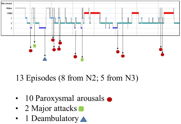
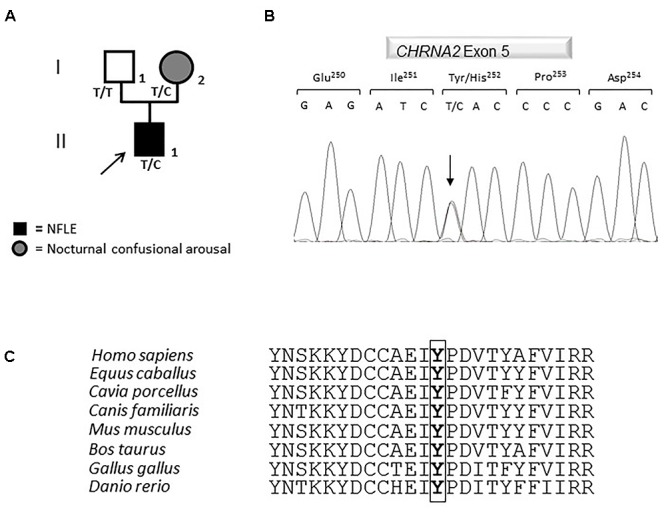
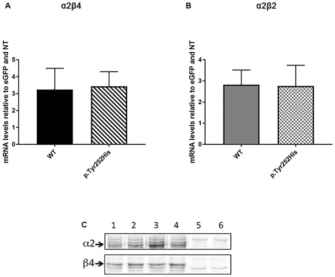
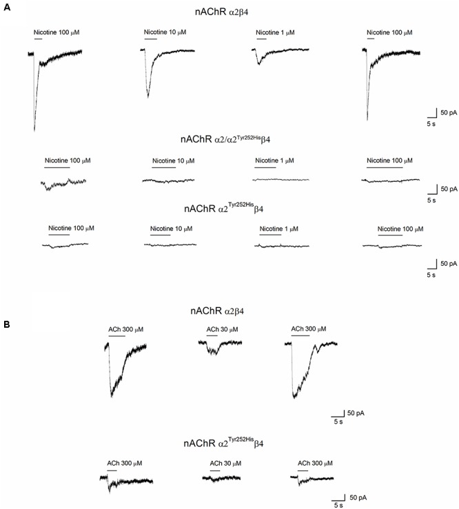
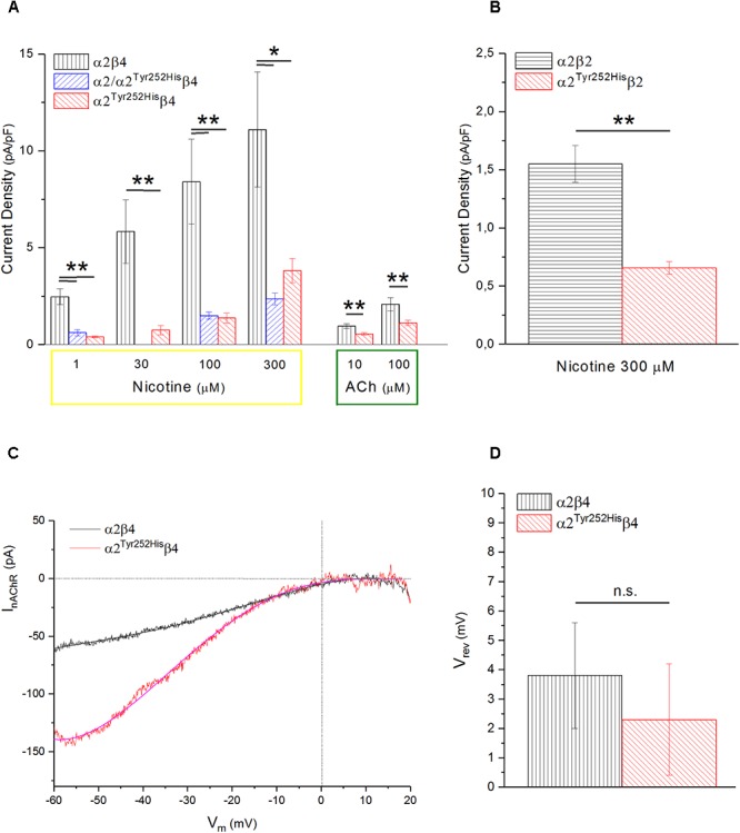
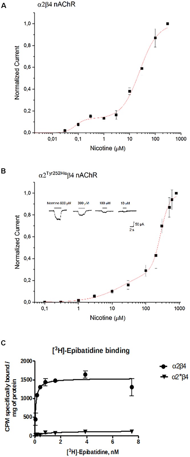
Mutations in genes coding for subunits of the neuronal nicotinic acetylcholine receptor (nAChR) have been involved in familial sleep-related hypermotor epilepsy (also named autosomal dominant nocturnal frontal lobe epilepsy, ADNFLE). Most of these mutations reside in CHRNA4 and CHRNB2 genes, coding for the α4 and β2 nAChR subunits, respectively. Two mutations with contrasting functional effects were also identified in the CHRNA2 gene coding for the α2 subunit. Here, we report the third mutation in the CHRNA2, found in a patient showing ADNFLE. The patient was examined by scalp EEG, contrast-enhanced brain magnetic resonance imaging (MRI), and nocturnal video-polysomnographic recording. All exons and the exon-intron boundaries of CHRNA2, CHRNA4, CHRNB2, CRH, KCNT1 were amplified and Sanger sequenced. In the proband, we found a c.754T>C (p.Tyr252His) missense mutation located in the N-terminal ligand-binding domain and inherited from the mother. Functional studies were performed by transient co-expression of α2 and α2 Tyr252His , with either β2 or β4, in human embryonic kidney (HEK293) cells. Equimolar amounts of subunits expression were obtained by using F2A-based multi-cistronic constructs encoding for the genes relative to the nAChR subunits of interest and for the enhanced green fluorescent protein. The mutation reduced the maximal currents by approximately 80% in response to saturating concentrations of nicotine in homo- and heterozygous form, in both the α2β4 and α2β2 nAChR subtypes. The effect was accompanied by a strong right-shift of the concentration-response to nicotine. Similar effects were observed using ACh. Negligible effects were produced by α2Tyr252His on the current reversal potential. Moreover, binding of (±)-[3H]Epibatidine revealed an approximately 10-fold decrease of both Kd and Bmax (bound ligand in saturating conditions), in cells expressing α2Tyr252His. The reduced Bmax and whole-cell currents were not caused by a decrease in mutant receptor expression, as minor effects were produced by α2Tyr252His on the level of transcripts and the membrane expression of α2β4 nAChR. Overall, these results suggest that α2Tyr252His strongly reduced the number of channels bound to the agonist, without significantly altering the overall channel expression. We conclude that mutations in CHRNA2 are more commonly linked to ADNFLE than previously thought, and may cause a loss-of-function phenotype.
Keywords: ADNFLE; ADSHE; frontal lobe epilepsy; genetics; nicotinic receptor; patch-clamp.
Figures
Similar articles
-
Nicotinic Receptors in Sleep-Related Hypermotor Epilepsy: Pathophysiology and Pharmacology.Brain Sci. 2020 Nov 25;10(12):907. doi: 10.3390/brainsci10120907. Brain Sci. 2020. PMID: 33255633 Free PMC article. Review.
-
Nocturnal frontal lobe epilepsy with paroxysmal arousals due to CHRNA2 loss of function.Neurology. 2015 Apr 14;84(15):1520-8. doi: 10.1212/WNL.0000000000001471. Epub 2015 Mar 13. Neurology. 2015. PMID: 25770198 Free PMC article.
-
Exclusion of linkage of nine neuronal nicotinic acetylcholine receptor subunit genes expressed in brain in autosomal dominant nocturnal frontal lobe epilepsy in four unrelated families.J Neurol. 2002 Aug;249(8):967-74. doi: 10.1007/s00415-002-0763-8. J Neurol. 2002. PMID: 12195439
-
A novel mutation of the nicotinic acetylcholine receptor gene CHRNA4 in sporadic nocturnal frontal lobe epilepsy.Epilepsy Res. 2009 Feb;83(2-3):152-6. doi: 10.1016/j.eplepsyres.2008.10.009. Epub 2008 Dec 5. Epilepsy Res. 2009. PMID: 19058950
-
[Generation of epilepsy animal model bearing a genetic abnormality identified in autosomal dominant nocturnal frontal lobe epilepsy (ADNFLE) of humans].Nihon Shinkei Seishin Yakurigaku Zasshi. 2010 Feb;30(1):9-14. Nihon Shinkei Seishin Yakurigaku Zasshi. 2010. PMID: 20297737 Review. Japanese.
Cited by
-
Nicotinic Receptors in Sleep-Related Hypermotor Epilepsy: Pathophysiology and Pharmacology.Brain Sci. 2020 Nov 25;10(12):907. doi: 10.3390/brainsci10120907. Brain Sci. 2020. PMID: 33255633 Free PMC article. Review.
-
Whole-Genome Sequencing Among Kazakhstani Children with Early-Onset Epilepsy Revealed New Gene Variants and Phenotypic Variability.Mol Neurobiol. 2023 Aug;60(8):4324-4335. doi: 10.1007/s12035-023-03346-3. Epub 2023 Apr 24. Mol Neurobiol. 2023. PMID: 37095367 Free PMC article.
-
Chromosome 15q BP4-BP5 Deletion in a Girl with Nocturnal Frontal Lobe Epilepsy, Migraine, Circumscribed Hypertrichosis, and Language Impairment.J Epilepsy Res. 2020 Dec 31;10(2):84-91. doi: 10.14581/jer.20014. eCollection 2020 Dec. J Epilepsy Res. 2020. PMID: 33659201 Free PMC article.
-
Cytisine Exerts an Anti-Epileptic Effect via α7nAChRs in a Rat Model of Temporal Lobe Epilepsy.Front Pharmacol. 2021 Jun 24;12:706225. doi: 10.3389/fphar.2021.706225. eCollection 2021. Front Pharmacol. 2021. PMID: 34248648 Free PMC article.
-
Genetic variations associated with pharmacoresistant epilepsy (Review).Mol Med Rep. 2020 Apr;21(4):1685-1701. doi: 10.3892/mmr.2020.10999. Epub 2020 Feb 24. Mol Med Rep. 2020. PMID: 32319641 Free PMC article. Review.
References
LinkOut - more resources
Full Text Sources