. 2019 Feb 12:12:34.
doi: 10.3389/fnmol.2019.00034. eCollection 2019.

Clinical Imaging of Choroid Plexus in Health and in Brain Disorders: A Mini-Review

Affiliations

Clinical Imaging of Choroid Plexus in Health and in Brain Disorders: A Mini-Review

Violaine Hubert et al. Front Mol Neurosci. .

Abstract

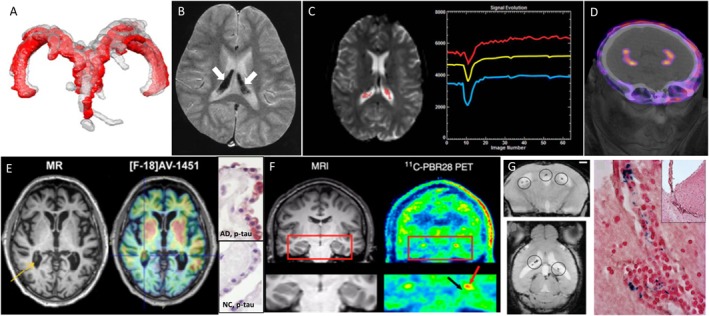
The choroid plexuses (ChPs) perform indispensable functions for the development, maintenance and functioning of the brain. Although they have gained considerable interest in the last years, their involvement in brain disorders is still largely unknown, notably because their deep location inside the brain hampers non-invasive investigations. Imaging tools have become instrumental to the diagnosis and pathophysiological study of neurological and neuropsychiatric diseases. This review summarizes the knowledge that has been gathered from the clinical imaging of ChPs in health and brain disorders not related to ChP pathologies. Results are discussed in the light of pre-clinical imaging studies. As seen in this review, to date, most clinical imaging studies of ChPs have used disease-free human subjects to demonstrate the value of different imaging biomarkers (ChP size, perfusion/permeability, glucose metabolism, inflammation), sometimes combined with the study of normal aging. Although very few studies have actually tested the value of ChP imaging biomarkers in patients with brain disorders, these pioneer studies identified ChP changes that are promising data for a better understanding and follow-up of diseases such as schizophrenia, epilepsy and Alzheimer's disease. Imaging of immune cell trafficking at the ChPs has remained limited to pre-clinical studies so far but has the potential to be translated in patients for example using MRI coupled with the injection of iron oxide nanoparticles. Future investigations should aim at confirming and extending these findings and at developing translational molecular imaging tools for bridging the gap between basic molecular and cellular neuroscience and clinical research.

Keywords: blood-CSF barrier; central nervous system; choroid plexus; imaging; inflammation; neurological disease.


Figures

FIGURE 1
FIGURE 1
Examples of different clinical imaging techniques available for investigating the involvement of ChPs in brain disorders. (A) 3D reconstruction of ChP located within lateral ventricles space [red, before lumbar puncture (LP), and gray, after LP] in a patient with idiopathic intracranial hypertension (Lublinsky et al., 2018); (B) ChP iron deposition (arrows on hypointense signals) detected by susceptibility-weighted MRI in a patient who had received both ultrasmall superparamagnetic particles of iron oxide (USPIOs) 2 years earlier and multiple blood transfusions since (Daldrup-Link, 2017); (C) signal-time curves extracted in the ChP from dynamic susceptibility contras-enhanced MRI data: the yellow curve represents the mean signal calculated from all the pixels in the ChP volume, while the blue and red curves represent, respectively, the curve with lowest and highest baseline. After first passage of the gadolinium bolus (signal drop), the choroidal signals visually exceed the baseline which indicates gadolinium chelate extravasation into the ChP stroma (Bouzerar et al., 2013); (D) PET/CT images showing the normal ChP uptake of [68Ga]-DOTA-E-[c(RGDfK)]2 targeting αvβ3 integrin (This research was originally published in JNM. Lopez-Rodriguez et al., 2016); (E) in vivo imaging using the Tau tracer [18F]AV-1451 showing high retention in AD patient and post-mortem ChP histopathology showing immunoreactivity in epithelial cells (pink color) with antibodies against pan-Tau in AD but not in normal control (NC) (Ikonomovic et al., 2016); (F) MRI and translocator protein (TSPO) labeling [11C]PBR28 PET images in a patient with left-sided temporal lobe epilepsy showing higher uptake in ipsilateral than in contralateral side in the ChP of lateral ventricles (red arrow) and hippocampus (black arrow) (This research was originally published in JNM. Hirvonen et al., 2012); (G) in vivo MRI using the very small particles of iron oxide (VSOP) and post-mortem Prussian Blue staining in a rat model of multiple sclerosis: VSOP (circles) was detected in the inflamed ChP at peak disease (Millward et al., 2013).
FIGURE 2
FIGURE 2
Schematic of different clinical imaging techniques available for investigating the involvement of ChPs in brain disorders. ChP volume and the presence of calcification may be assessed through conventional imaging techniques such as CT and MRI. CSF production may be evaluated with phase-sensitive MRI sequences. ChP capillary perfusion and permeability may be quantified with MRI using dynamic imaging coupled to small molecular weight contrast agents such as gadolinium chelates. Nuclear medicine techniques such as PET give access to: (i) the occurrence of receptors on stromal or epithelial cells (both at the basal or at the apical side) and (ii) epithelial cell metabolism, thanks to dedicated radiotracers. All of these morphological and functional features present a progressive degradation throughout the aging process which might get aggravated in neurological diseases. In addition, in pathological conditions, the extravasation of small molecular weight contrast agents into CSF may allow to image blood-CSF barrier disruption. In inflammatory conditions, reticuloendothelial MRI contrast agents such as iron oxide nanoparticles may get access to the ChP stroma and accumulate into endothelial, stromal, and/or epithelial cells. Alternatively, activation of stromal immune cells (whether resident or infiltrating) may be observed with TSPO-targeted PET radiotracers. Finally, the presence of aggregated proteins due to proteinopathies might be detected with dedicated PET tracers, although this is still debated due to “off-target” binding. ChPs, choroid plexuses; CSF, cerebrospinal fluid; BCSFB, blood-cerebrospinal fluid barrier; MDR, multidrug-resistant; CT, computed tomography; MRI, magnetic resonance imaging; PET, positron emission tomography; TSPO, translocator protein 18 kDa (this figure was prepared with Servier Medical Art).

Similar articles

Cited by

References

    1. Alicioglu B., Yilmaz G., Tosun O., Bulakbasi N. (2017). Diffusion-weighted magnetic resonance imaging in the assessment of choroid plexus aging. Neuroradiol. J. 30 490–495. 10.1177/1971400917714280 - DOI - PMC - PubMed
    1. Anderson I. M., Clark L., Elliott R., Kulkarni B., Williams S. R., Deakin J. F. (2002). 5-HT(2C) receptor activation by m-chlorophenylpiperazine detected in humans with fMRI. Neuroreport 13 1547–1551. 10.1097/00001756-200208270-00012 - DOI - PubMed
    1. Artzi M., Aizenstein O., Abramovitch R., Bashat D. B. (2013). MRI multiparametric hemodynamic characterization of the normal brain. Neuroscience 240 269–276. 10.1016/j.neuroscience.2013.03.004 - DOI - PubMed
    1. Azuma M., Hirai T., Kadota Y., Khant Z. A., Hattori Y., Kitajima M., et al. (2018). Circumventricular organs of human brain visualized on post-contrast 3D fluid-attenuated inversion recovery imaging. Neuroradiology 10.1007/s00234-018-2023-3 [Epub ahead of print]. - DOI - PubMed
    1. Batra A., Latour L. L., Ruetzler C. A., Hallenbeck J. M., Spatz M., Warach S., et al. (2010). Increased plasma and tissue MMP levels are associated with BCSFB and BBB disruption evident on post-contrast FLAIR after experimental stroke. J. Cereb. Blood Flow Metab. 30 1188–1199. 10.1038/jcbfm.2010.1 - DOI - PMC - PubMed