The Ever-Growing Puzzle of Asynchronous Release
- PMID: 30809127
- PMCID: PMC6379310
- DOI: 10.3389/fncel.2019.00028
The Ever-Growing Puzzle of Asynchronous Release
Abstract
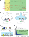
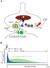
Invasion of an action potential (AP) to presynaptic terminals triggers calcium dependent vesicle fusion in a relatively short time window, about a millisecond, after the onset of the AP. This allows fast and precise information transfer from neuron to neuron by means of synaptic transmission and phasic mediator release. However, at some synapses a single AP or a short burst of APs can generate delayed or asynchronous synaptic release lasting for tens or hundreds of milliseconds. Understanding the mechanisms underlying asynchronous release (AR) is important, since AR can better recruit extrasynaptic metabotropic receptors and maintain a high level of neurotransmitter in the extracellular space for a substantially longer period of time after presynaptic activity. Over the last decade substantial work has been done to identify the presynaptic calcium sensor that may be involved in AR. Several models have been suggested which may explain the long lasting presynaptic calcium elevation a prerequisite for prolonged delayed release. However, the presynaptic mechanisms underlying asynchronous vesicle release are still not well understood. In this review article, we provide an overview of the current state of knowledge on the molecular components involved in delayed vesicle fusion and in the maintenance of sufficient calcium concentration to trigger AR. In addition, we discuss possible alternative models that may explain intraterminal calcium dynamics underlying AR.
Keywords: calcium; calcium extrusion; presynaptic; synaptic release; synaptotagmins.
Figures


Similar articles
-
Synaptotagmin 7 Mediates Both Facilitation and Asynchronous Release at Granule Cell Synapses.J Neurosci. 2018 Mar 28;38(13):3240-3251. doi: 10.1523/JNEUROSCI.3207-17.2018. J Neurosci. 2018. PMID: 29593071 Free PMC article.
-
Synaptotagmins 3 and 7 mediate the majority of asynchronous release from synapses in the cerebellum and hippocampus.Cell Rep. 2024 Aug 27;43(8):114595. doi: 10.1016/j.celrep.2024.114595. Epub 2024 Aug 6. Cell Rep. 2024. PMID: 39116209 Free PMC article.
-
Vesicle dynamics: how synaptic proteins regulate different modes of neurotransmission.J Neurochem. 2013 Jul;126(2):146-54. doi: 10.1111/jnc.12245. Epub 2013 Apr 23. J Neurochem. 2013. PMID: 23517499 Review.
-
Neurotransmitter Release Can Be Stabilized by a Mechanism That Prevents Voltage Changes Near the End of Action Potentials from Affecting Calcium Currents.J Neurosci. 2016 Nov 9;36(45):11559-11572. doi: 10.1523/JNEUROSCI.0066-16.2016. J Neurosci. 2016. PMID: 27911759 Free PMC article.
-
[Research progress of synaptic vesicle recycling].Sheng Li Xue Bao. 2015 Dec 25;67(6):545-60. Sheng Li Xue Bao. 2015. PMID: 26701630 Review. Chinese.
Cited by
-
The Core Complex of the Ca2+-Triggered Presynaptic Fusion Machinery.J Mol Biol. 2023 Jan 15;435(1):167853. doi: 10.1016/j.jmb.2022.167853. Epub 2022 Oct 13. J Mol Biol. 2023. PMID: 36243149 Free PMC article. Review.
-
Function of Drosophila Synaptotagmins in membrane trafficking at synapses.Cell Mol Life Sci. 2021 May;78(9):4335-4364. doi: 10.1007/s00018-021-03788-9. Epub 2021 Feb 22. Cell Mol Life Sci. 2021. PMID: 33619613 Free PMC article. Review.
-
Control of synaptic vesicle release probability via VAMP4 targeting to endolysosomes.Sci Adv. 2021 Apr 30;7(18):eabf3873. doi: 10.1126/sciadv.abf3873. Print 2021 Apr. Sci Adv. 2021. PMID: 33931449 Free PMC article.
-
Complexin has a dual synaptic function as checkpoint protein in vesicle priming and as a promoter of vesicle fusion.Proc Natl Acad Sci U S A. 2024 Apr 9;121(15):e2320505121. doi: 10.1073/pnas.2320505121. Epub 2024 Apr 3. Proc Natl Acad Sci U S A. 2024. PMID: 38568977 Free PMC article.
-
Nano-Organization at the Synapse: Segregation of Distinct Forms of Neurotransmission.Front Synaptic Neurosci. 2021 Dec 22;13:796498. doi: 10.3389/fnsyn.2021.796498. eCollection 2021. Front Synaptic Neurosci. 2021. PMID: 35002671 Free PMC article. Review.
References
Publication types
LinkOut - more resources
Full Text Sources
Research Materials
Miscellaneous

