. 2019 Feb 12:10:213.
doi: 10.3389/fimmu.2019.00213. eCollection 2019.

Increased Regulatory T-Cell Activity and Enhanced T-Cell Homeostatic Signaling in Slow Progressing HIV-infected Children

Affiliations

Increased Regulatory T-Cell Activity and Enhanced T-Cell Homeostatic Signaling in Slow Progressing HIV-infected Children

Julia Roider et al. Front Immunol. .

Abstract

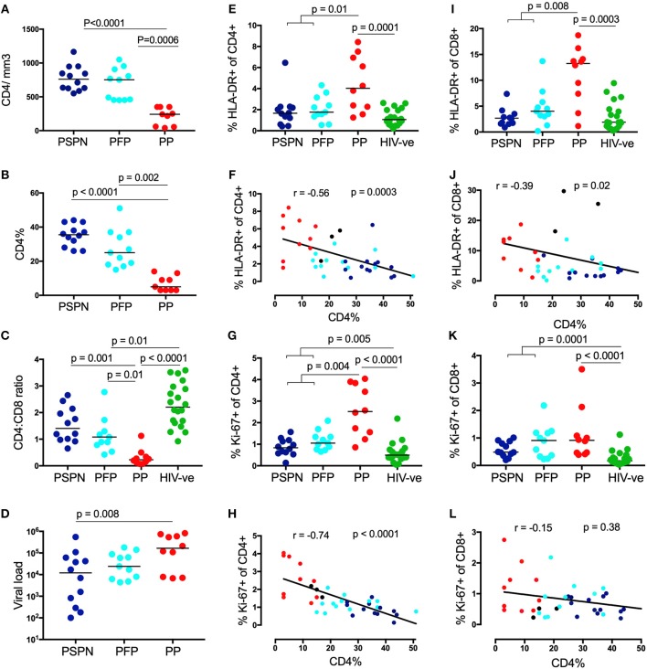
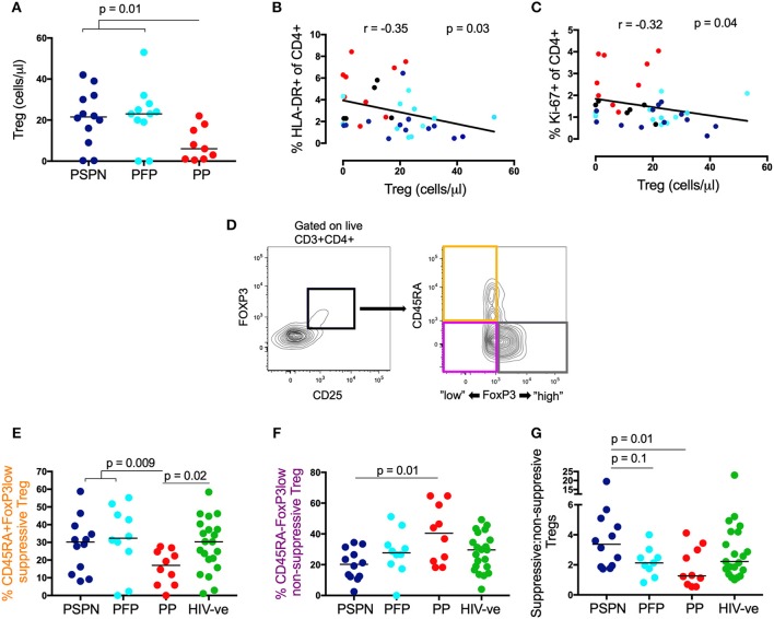
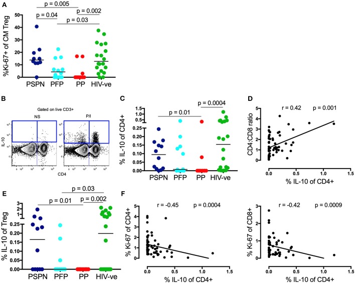
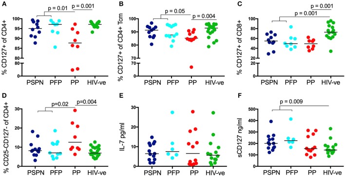
Pediatric slow progressors (PSP) are rare ART-naïve, HIV-infected children who maintain high CD4 T-cell counts and low immune activation despite persistently high viral loads. Using a well-defined cohort of PSP, we investigated the role of regulatory T-cells (TREG) and of IL-7 homeostatic signaling in maintaining normal-for-age CD4 counts in these individuals. Compared to children with progressive disease, PSP had greater absolute numbers of TREG, skewed toward functionally suppressive phenotypes. As with immune activation, overall T-cell proliferation was lower in PSP, but was uniquely higher in central memory TREG (CM TREG), indicating active engagement of this subset. Furthermore, PSP secreted higher levels of the immunosuppressive cytokine IL-10 than children who progressed. The frequency of suppressive TREG, CM TREG proliferation, and IL-10 production were all lower in PSP who go on to progress at a later time-point, supporting the importance of an active TREG response in preventing disease progression. In addition, we find that IL-7 homeostatic signaling is enhanced in PSP, both through preserved surface IL-7receptor (CD127) expression on central memory T-cells and increased plasma levels of soluble IL-7receptor, which enhances the bioactivity of IL-7. Combined analysis, using a LASSO modeling approach, indicates that both TREG activity and homeostatic T-cell signaling make independent contributions to the preservation of CD4 T-cells in HIV-infected children. Together, these data demonstrate that maintenance of normal-for-age CD4 counts in PSP is an active process, which requires both suppression of immune activation through functional TREG, and enhanced T-cell homeostatic signaling.

Keywords: homeostatic signaling; IL-10; IL-7; immune activation (IA); immune regulation; pediatric HIV-infection; pediatric slow progression; regulatory T cells (Treg).


Figures

Figure 1
Figure 1
Pediatric slow progressors show low immune activation preferentially on CD4 T-cells. CD4 count absolute (A), CD4 percentage (B), viral load (C) in three pediatric groups: Pediatric slow progressors (PSP; n = 23) were defined here as treatment naïve, aged >5 years with CD4 counts >450/mm3 at the time point the experiments were performed. According to the longitudinal follow up we separated “pediatric future progressors” (PFP; n = 11; light blue) who would go onto treatment from true pediatric slow progressors, who stayed treatment naïve during the follow-up (PSPN; n = 12; dark blue). Pediatric progressors (PP; n = 10; red) were defined as treatment naïve, aged >5 years with CD4 counts <350/mm3. For CD4:CD8 ratio (D) age-matched uninfected controls (n = 20; green) were included as control group. (E) Frequency of HLA expression and its correlation with CD4 percentage (F) on CD4 T-cells within the 4 pediatric groups. (G,H), same as (E,F) but for Ki-67 expression. (I–L) same as (E–H) but of CD8 T-cells. For scatterplots, median, and interquartile range are shown. Kruskal-Wallis test was performed and corrected for multiple comparisons. Only significant p-values (<0.05) are given. For correlations, Spearman ranks test was used. For correlations with CD4 percentage, all HIV-infected infants were used including those not fulfilling criteria for PSP nor PP (black dots; n = 3; CD4 count >350 and <450/mm3).
Figure 2
Figure 2
Suppressive regulatory T-cells are increased in pediatric slow progressors. (A) Absolute TREG count/μl (gated on CD25+FoxP3+ CD4 T-cells) within the different pediatric groups: PSPN (dark blue; n = 12), PFP (light blue; n = 11), PP (red; n = 10) and (B) its correlation with HLA-DR and Ki-67 (C) on CD4 T-cells. For correlations all HIV-infected children were used including those not fulfilling criteria for PSP nor PP (black dots; n = 3; CD4 count >350 and <450/mm3). (D) Gating strategy for three functional TREG (gated of CD25+FoxP3+ CD4 T-cells) memory subsets (39): (i) “activated”-suppressive TREG (CD45RAFoxP3high; gray); (ii) “resting”-suppressive TREG (CD45RA+FoxP3low; orange) and cytokine-producing non-suppressive TREG (CD45RAFoxP3low; pink). (E) Same groups as (A) but including uninfected controls (green; n = 20) and showing the frequency of “resting”- suppressive TREG (CD45RA+FoxP3low) and cytokine-producing non-suppressive TREG (CD45RAFoxP3low; F). (G) Same as (E,F) but showing the ratio of all suppressive TREG (CD45RA+FoxP3low and CD45RAFoxP3high; orange and gray population, respectively) to non-suppressive TREG (CD45RAFoxP3low; pink population) within the pediatric groups. For scatterplots, median, and interquartile range are shown. Kruskal-Wallis test was performed and corrected for multiple comparisons. For correlations, Spearman ranks test was used.
Figure 3
Figure 3
Memory TREG proliferation and IL-10 secretion are increased in pediatric slow progressors. (A) Frequency of Ki-67+ve “central memory” TREG (gated on CD4+CD25+FoxP3+CD45RACCR7+) within the pediatric groups: PSPN (dark blue; n = 12), PFP (light blue; n = 11), PP (red; n = 10), and uninfected controls (green; n = 20). (B) Exemplary FACS plot of one PSPN study subject showing CD4 (x-axis) vs. IL-10 (y-axis) staining (left: unstimulated control, right: stimulated with PMA/Ionomycin). (C) Same groups as (A) but showing IL-10 production of CD4 T-cells as measured by intracellular staining assay after 5 h stimulation with PMA/ Ionomycin and (D) its correlation with CD4:CD8 ratio within the whole pediatric cohort. (E) Same as (C) but of TREG. (F) Same as (D) but correlating IL-10 production of CD4 T-cells with Ki-67 expression on CD4 (left) and CD8 (right) T-cells. For correlations, all data available from pediatric subjects was used, including those not fitting criteria for PSP nor PP. For scatterplots, median, and interquartile range are shown. For correlations, Spearman ranks test was used. Kruskal-Wallis test was performed and corrected for multiple comparisons.
Figure 4
Figure 4
Increased IL-7/sIL-7 receptor in pediatric slow progressors. Frequency of IL-7R (CD127) expression on CD4 (A) and on central memory CD4 (B) T-cells in PSPN (dark blue; n = 10), PFP (light blue; n = 8), PP (red; n = 8), and uninfected pediatric controls (green; n = 15). (C) Same groups as before but showing frequency of IL-7R expression on CD8 T-cells and (D) frequency of CD4 T-cells double-negative for IL-2R (CD25) and IL-7R (CD127). (E) Ex-vivo plasma levels of IL-7 in pg/ml in PSPN (n = 13, dark blue), PFP (n = 6, light blue), PP (n = 12; red), and pediatric uninfected controls (n = 14; green). (F) Same as in (E) but showing plasma levels of IL-7R (sCD127) in ng/ml in the different groups. For scatterplots, median, and interquartile range are shown. Kruskal-Wallis test was performed and corrected for multiple comparisons.

Similar articles

Cited by

References

    1. Goulder PJ, Lewin SR, Leitman EM. Paediatric HIV infection: the potential for cure. Nat Rev Immunol. (2016) 16:259–71. 10.1038/nri.2016.19 - DOI - PMC - PubMed
    1. Blanche S, Newell ML, Mayaux MJ, Dunn DT, Teglas JP, Rouzioux C, et al. . Morbidity and mortality in European children vertically infected by HIV-1. The french pediatric HIV infection study group and european collaborative study. J Acquir Immune Defic Syndr Hum Retrovirol. (1997) 14:442–50 - PubMed
    1. Paul ME, Mao C, Charurat M, Serchuck L, Foca M, Hayani K, et al. . Predictors of immunologic long-term nonprogression in HIV-infected children: implications for initiating therapy. J Allergy Clin Immunol. (2005) 115:848–55. 10.1016/j.jaci.2004.11.054 - DOI - PubMed
    1. Ssewanyana I, Elrefaei M, Dorsey G, Ruel T, Jones NG, Gasasira A, et al. . Profile of T cell immune responses in HIV-infected children from Uganda. J Infect Dis. (2007) 196:1667–70. 10.1086/522013 - DOI - PubMed
    1. Ananworanich J, Apornpong T, Kosalaraksa P, Jaimulwong T, Hansudewechakul R, Pancharoen C, et al. . Characteristics of lymphocyte subsets in HIV-infected, long-term nonprogressor, and healthy Asian children through 12 years of age. J Allergy Clin Immunol. (2010) 126:1294–1301 e1210. 10.1016/j.jaci.2010.09.038 - DOI - PMC - PubMed

Publication types

MeSH terms