. 2019 Jan 28:2019:2746521.
doi: 10.1155/2019/2746521. eCollection 2019.

Excessive Oxidative Stress Contributes to Increased Acute ER Stress Kidney Injury in Aged Mice

Affiliations

Excessive Oxidative Stress Contributes to Increased Acute ER Stress Kidney Injury in Aged Mice

Xiaoyan Liu et al. Oxid Med Cell Longev. .

Abstract

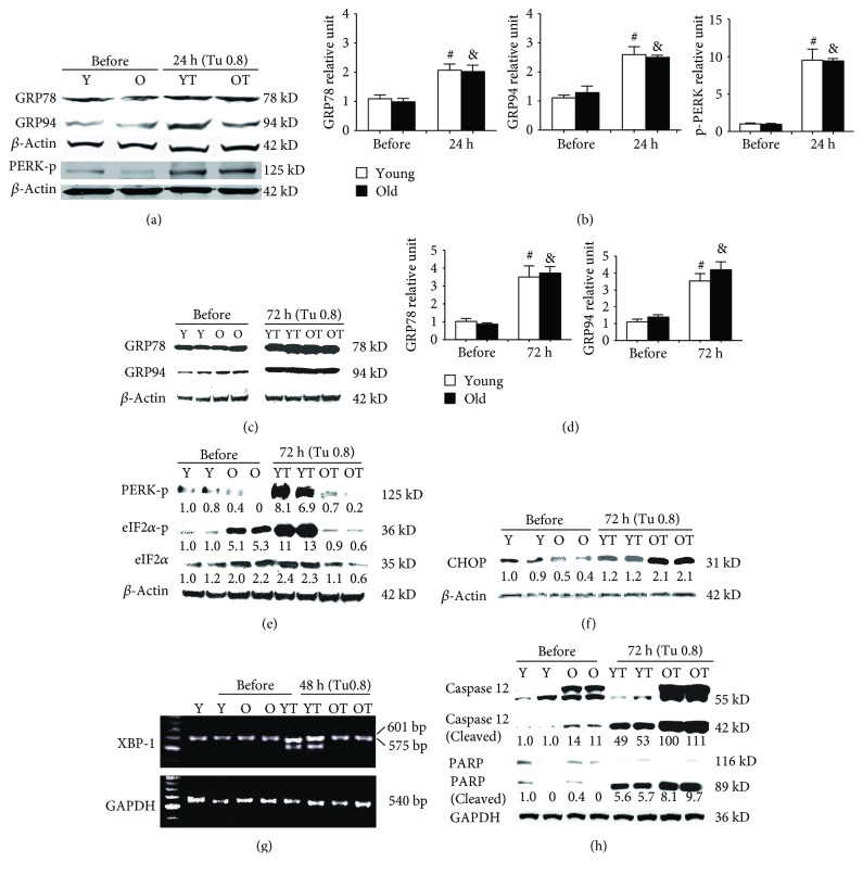
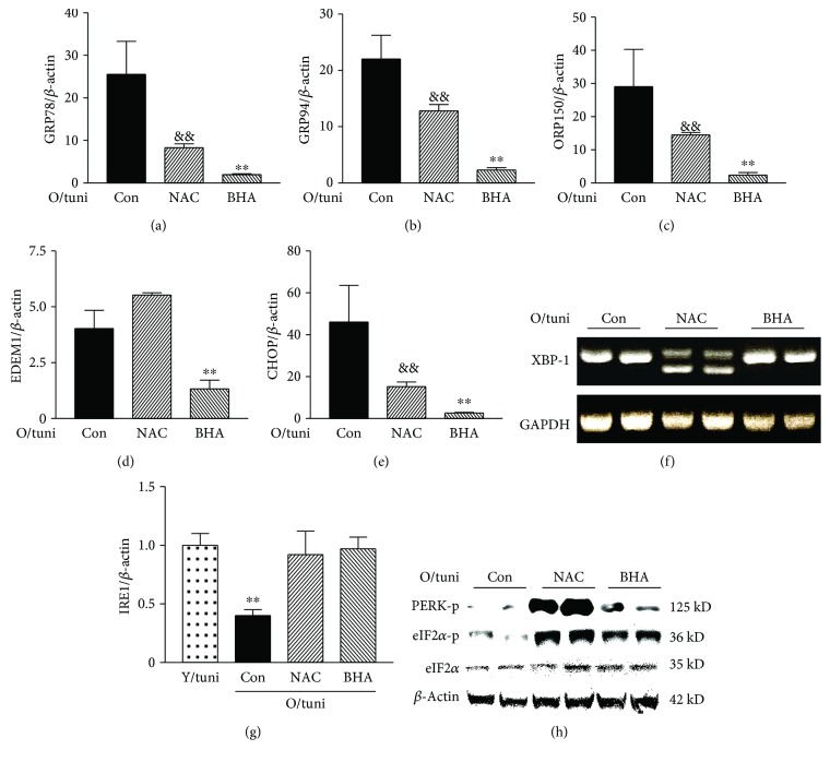
The aged kidney is susceptible to acute injury due presumably to its decreased ability to handle additional challenges, such as endoplasmic reticulum (ER) stress. This was tested by giving tunicamycin, an ER stress inducer, to either old or young mice. Injection of high dose caused renal failure in old mice, not in young mice. Moreover, injection of low dose resulted in severe renal damage in old mice, confirming the increased susceptibility of aged kidney to ER stress. There existed an abnormality in ER stress response kinetics in aged kidney, characterized by a loss of XBP-1 splicing and decreased PERK-eIF2α phosphorylation at late time point. The presence of excessive oxidative stress in aged kidney may play a role since high levels of oxidation increased ER stress-induced cell death and decreased IRE1 levels and XBP-1 splicing. Importantly, treatment with antioxidants protected old mice from kidney injury and normalized IRE1 and XBP-1 responses. Furthermore, older mice (6 months old) transgenic with antioxidative stress AGER1 were protected from ER stress-induced kidney injury. In conclusion, the decreased ability to handle ER stress, partly due to the presence of excessive oxidative stress, may contribute to increased susceptibility of the aging kidney to acute injury.


Figures

Figure 1
Figure 1
Differences in kidney function and renal lesions between old and young mice after high dose of tunicamycin injury (0.8 μg/g BW, n = 8/age group). (a, b) Increased BUN and Scr levels in old mice after high dose of tunicamycin injection: serum samples were obtained from 18–22-month- or 3–6-month-old mice prior to and after 72 hours of high dose of tunicamycin injection. (c, d) Representative kidney sections from (c) young and (d) old mice without receiving tunicamycin seem normal. (e, g) Extensive tubular vacuolation was present in young mice at 72 hours of high dose tunicamycin injury (PAS, (e) 200x, and (g) 400x). The lesions were localized in proximal tubules (red arrow) while the descending tubules extended from the injured proximal tubule were relatively normal as indicated by a (e) black arrow. (g) Nuclear pyknosis (condensation) and fragmentation were widely present (arrows). (f) Large vacuoles were less common in the kidneys of tunicamycin-treated old mice (200x). (f) However, the cellular damage was more severe with detachment of the whole segment of proximal tubular cells from the basement membrane in old mice (arrows). (h) Higher power magnification (400x) showed that cells in the injured tubules in aging kidney contained many small or fine vacuoles. Nuclear damages were also prominent. (i) Morphometry analysis revealed that tubular damage occurred nearly in all proximal tubules in old mice after high dose of tunicamycin injection. More apoptotic cell death in aging kidney after high dose of tunicamycin injury: apoptotic TUNEL staining was performed in kidney sections obtained from (l) young or (m) old mice at 72 hours of high-dose tunicamycin injection. Kidneys without tunicamycin treatment for (j) young and (k) old mice were as controls (400x). Nuclei with brown or black staining (DAB) were apoptotic cells (arrows). (n) More apoptotic cell counts in the kidneys from tunicamycin-treated old mice. p < 0.05 vs. young mice. ∗∗ p < 0.01 vs. the levels before tunicamycin injection. Data was expressed as mean ± SD. Scale bar = 50 μm.
Figure 2
Figure 2
Differences in mRNA levels of UPR-related genes between the kidneys of old and young mice after high dose of tunicamycin injury: renal cortex RNA was obtained from young and old mice at baseline, 24 hours, and 72 hours after high dose of tunicamycin (0.8 μg/g) injection. The levels of GRP78, GRP94, ORP150, EDEM1, and CHOP mRNAs were measured by real-time PCR, and data was expressed as the ratio after dividing with β-actin mRNA levels at the same sample. & p < 0.05 vs. young mice at baseline. # p < 0.05 vs. young mice at 24 hours. p < 0.05 vs. young mice at 72 hours.
Figure 3
Figure 3
Abnormalities in UPR in the kidneys of old mice: renal cortex RNA was obtained from young and old mice at baseline, 24 hours, 48 hours, and 72 hours after high dose of tunicamycin (0.8 μg/g) injection. The levels of GRP78, GRP94, phosphorylated PERK (PERK-p), phosphorylated eIF2α (eIF2α-p), total eIF2α, CHOP, caspase 12, and PARP were measured in eight animals from each group by Western blots. XBP-1 and GAPDH mRNA expression in kidneys at baseline and 48 hours after tunicamycin injection was determined by RT-PCR (n = 6). Results from two representative animals of baseline and tunicamycin-treated young and old mice were shown. The intensity of each blot band was quantitated using a densitometer. Data from the untreated kidneys of young mice was arbitrarily defined as 1 after correcting with the intensity of the individual β-actin band of the same sample. Lanes Y (baseline) and YT (tunicamycin treated) were samples from young mice. Lanes O (baseline) and OT (tunicamycin treated) were samples from old mice. Both GRP78 and GRP94 were significantly increased in the kidneys of both young and old mice at 24 (a, b) and 72 hours (c, d) after tunicamycin treatment. No differences were found between young and old mice. At baseline, phosphorylated PERK levels were low (a, e). The levels were significantly elevated in both old and young mice 24 hours after high dose of tunicamycin injection (a). Phosphorylated PERK remained high in young mice but was largely lost in old mice at 72 hours (e). Subsequently, at 72 hours, phosphorylated eIF2α was increased in young mice while it was decreased in old mice (e). High-dose tunicamycin treatment was associated with more increased kidney CHOP protein levels in old mice (f). XBP-1 mRNA splicing occurred only in the kidneys of young mice after treatment (g). Cleaved caspase 12 levels were increased in old mice at baseline and were further increased after tunicamycin treatment (h). The increase in cleaved PARP was also more robust in old mice (h). # p < 0.05 vs. young mice at baseline. & p < 0.05 vs. old mice at baseline.
Figure 4
Figure 4
ER stress-induced severe oxidative stress in the kidneys of old mice is prevented by antioxidants. Renal tissue was obtained from young and old mice at baseline, 72 hours after tunicamycin (0.8 μg/g) injection and old mice pretreatment with BHA for 7 days or NAC 24 hours before tunicamycin injection. ER stress caused elevation in MDA, oxidized protein, and AGEs and decreased reducing glutathione (GSH)/glutathione disulfide (GSSG). (a) Levels of MDA, (b) levels of oxidized protein, (c) levels of AGEs, and (d) the ratio of GSH and GSSG. Y: young mice; O: old mice; Y/tuni: young mice with tunicamycin injury; O/tuni: old mice with tunicamycin injury: NAC; old mice treated with NAC; BHA: old mice treated with BHA. # p < 0.05 and ## p < 0.01 vs. young mice at baseline; ∧∧ p < 0.01 vs. old mice at baseline; p < 0.05 and ∗∗ p < 0.01 vs. old mice that received tunicamycin; and && p < 0.01 vs. old mice treated with NAC.
Figure 5
Figure 5
Prevention of ER stress renal injury in old mice by antioxidants. NAC or BHA treatment blocked high dose of tunicamycin-induced elevation (0.8 μg/g) of BUN (a) and Scr (b). Histologically, while severe renal injury characterized by sloughing of tubular cells (arrow) was frequently seen in old mice that received tunicamycin alone (c), NAC treatment resulted in a partial protection against tunicamycin-induced renal injury in old mice (d). There is no tubular cell sloughing, but vacuolation and nuclear damage (arrow) were seen in NAC-treated mice (d). BHA treatment gave a nearly complete protection, and renal histology was essentially normal in this group (e). Morphometry quantitation of the injured area in the renal cortex revealed a significant reduction in ER stress renal injury by antioxidants, and the effect was especially prominent by BHA (f). Counting the number of TUNEL-positive cells in kidney sections showed that BHA and NAC decreased tunicamycin-induced apoptotic cell death (g). ∗∗ p < 0.01 vs. old mice that received tunicamycin alone (O/tu) and ## p < 0.01 vs. NAC-treated mice. Scale bar = 50 μm.
Figure 6
Figure 6
Antioxidant treatment corrects UPR dysregulation in old mice. RNA was collected from the kidneys of controls that received tunicamycin alone (0.8 μg/g) or treated with NAC or BHA before tunicamycin injection (n = 6/group). mRNA levels of GRP78 (a), GRP94 (b), OPR150 (c), EDEM1 (d), and CHOP (e) in the kidneys at 72 hours after tunicamycin injection were determined by real-time PCR and corrected for β-actin mRNA levels. O/tuni: old mice challenged with high dose of tunicamycin. && p < 0.01 vs. old mice treated with tunicamycin alone (Con). ∗∗ p < 0.01 vs. mice pretreated with NAC. XBP-1 spicing (f). Regular PCR was performed in the kidneys from tunicamycin alone (Con) and NAC- and BHA-treated mice at 48 hours after tunicamycin injection (n = 6/group). Representative gels from two mice of each group were shown. The loss of XBP-1 splicing in tunicamycin alone (Con) mice reappears in NAC-treated mice. IRE1 mRNA levels (g). ∗∗ p < 0.01 vs. young mice treated with high dose of tunicamycin or old mice pretreated with NAC or BHA. The levels of phosphorylated PERK, phosphorylated eIF2α, and total eIF2α in the kidneys from control and NAC- and BHA-treated old mice (h). Renal protein was obtained from these mice 72 hours after tunicamycin injection (n = 6/group). Representative gel shows two samples from each group. The levels of phosphorylated PERK and eIF2α were highest in the NAC-treated group. Phosphorylated eIF2α was also visibly higher in the BHA group than in the control group.

Similar articles

Cited by

References

    1. Brunnler T., Drey M., Dirrigl G., et al. The oldest old in the emergency department: impact of renal function. The Journal of Nutrition, Health & Aging. 2016;20(10):1045–1050. doi: 10.1007/s12603-016-0731-0. - DOI - PubMed
    1. Liaño F., Pascual J., The Madrid Acute Renal Failure Study Group Epidemiology of acute renal failure: a prospective, multicenter, community-based study. Kidney International. 1996;50(3):811–818. doi: 10.1038/ki.1996.380. - DOI - PubMed
    1. Chen G., Bridenbaugh E. A., Akintola A. D., et al. Increased susceptibility of aging kidney to ischemic injury: identification of candidate genes changed during aging, but corrected by caloric restriction. American Journal of Physiology-Renal Physiology. 2007;293(4):F1272–F1281. doi: 10.1152/ajprenal.00138.2007. - DOI - PMC - PubMed
    1. Adler S., Huang H., Wolin M. S., Kaminski P. M. Oxidant stress leads to impaired regulation of renal cortical oxygen consumption by nitric oxide in the aging kidney. Journal of the American Society of Nephrology. 2004;15(1):52–60. doi: 10.1097/01.ASN.0000101032.21097.C5. - DOI - PubMed
    1. Anderson S., Brenner B. M. The aging kidney: structure, function, mechanisms, and therapeutic implications. Journal of the American Geriatrics Society. 1987;35(6):590–593. doi: 10.1111/j.1532-5415.1987.tb01407.x. - DOI - PubMed

MeSH terms