

Single-particle tracking discloses binding-mediated rocking diffusion of rod-shaped biological particles on lipid membranes
- PMID: 30809350
- PMCID: PMC6354740
- DOI: 10.1039/c8sc04033h
Single-particle tracking discloses binding-mediated rocking diffusion of rod-shaped biological particles on lipid membranes
Abstract
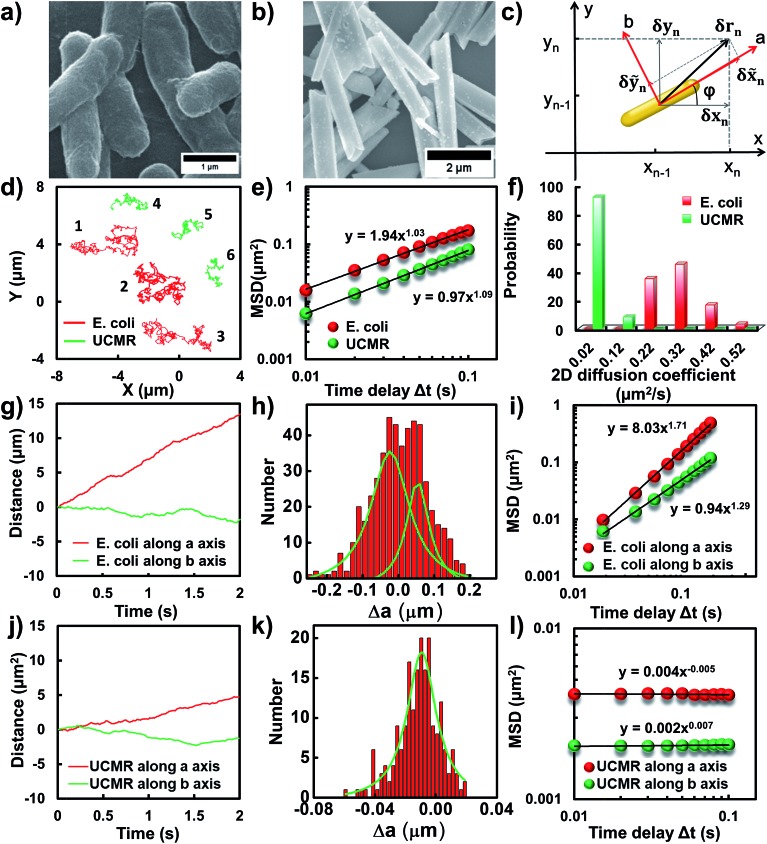
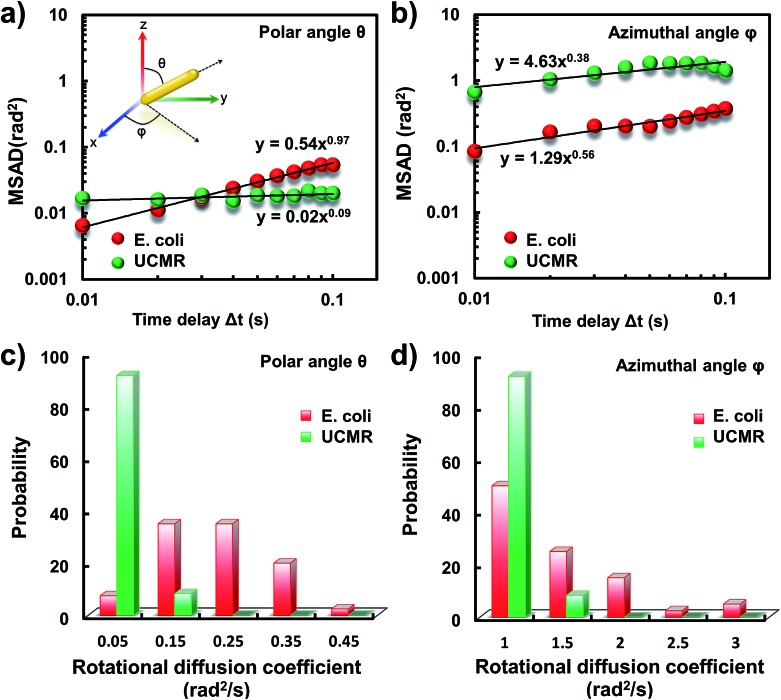
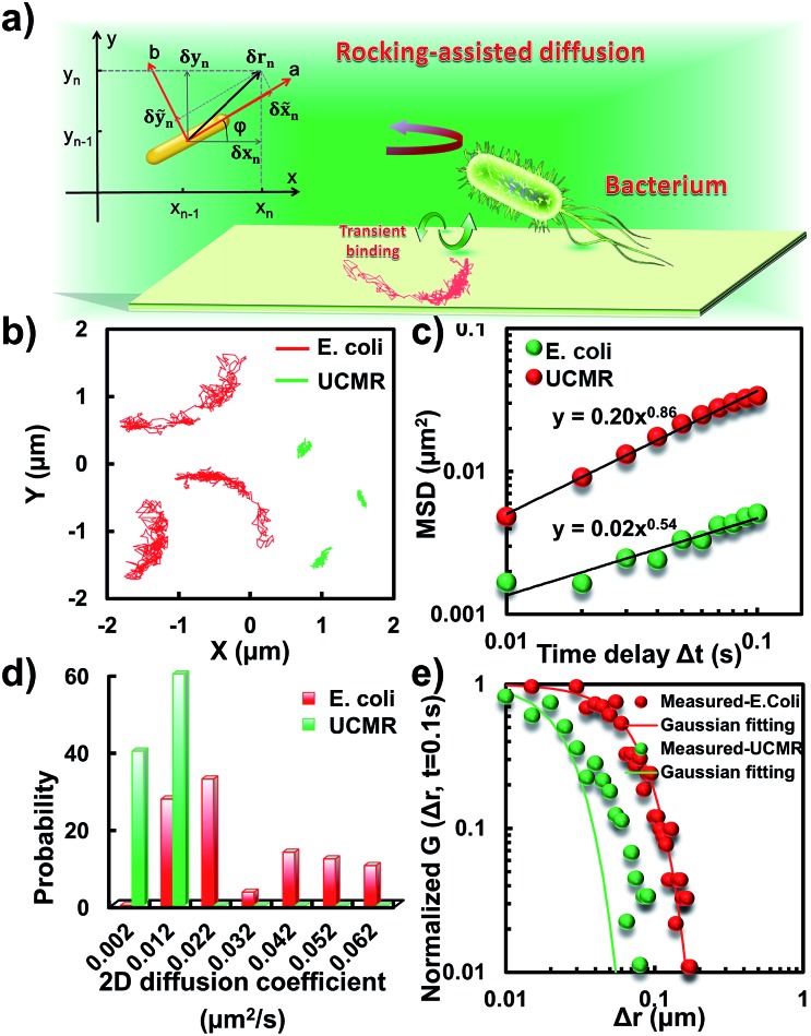
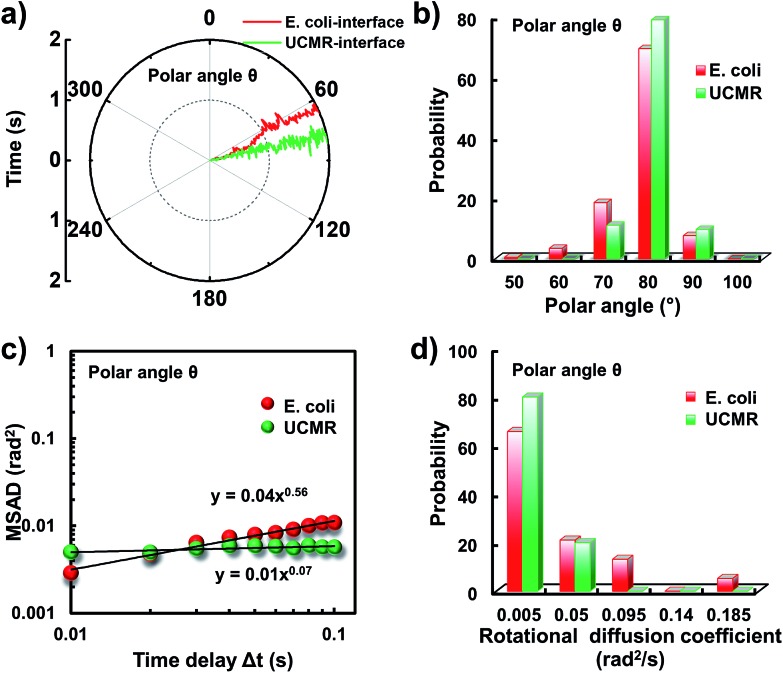
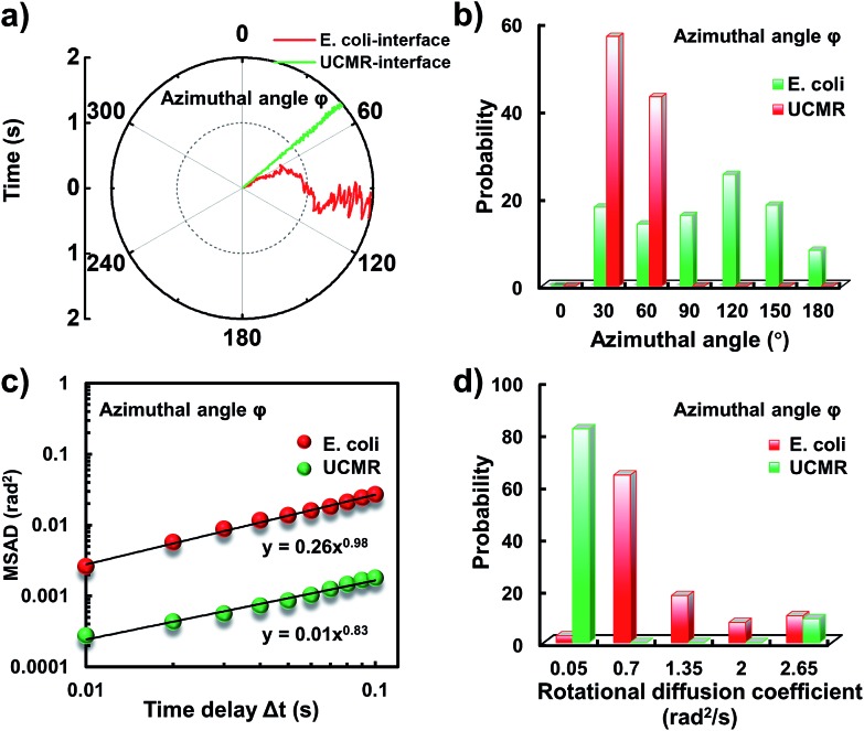
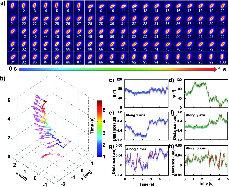
It has been demonstrated that rod-shaped particles can achieve a high translocation efficiency for gene and drug delivery in biological samples. Previous theoretical calculations also confirmed that rod-shaped particles display higher diffusivity than their spherical counterparts in biological porous media. Understanding the diffusion dynamics of biological and non-biological rod-shaped particles in biological solutions as well as close to the lipid membrane is therefore fundamentally significant for the rational design of efficient cargos. With dark-field optical microscopy, the translational and three-dimensional (3D) orientational diffusion dynamics of individual rod-shaped particles (i.e., E. coli and upconversion microrods, UCMRs) in phosphate buffered saline (PBS) and on the lipid membrane are tracked at the single-particle level. In the buffer solution, faster rotation of E. coli in the z direction was observed even though its dynamics in the x-y plane is comparable with that of UCMRs. Interestingly, on the lipid membrane, distinct from the confined motion of UCMRs, anomalous rocking diffusion was observed, which might facilitate the subsequent survey of stronger association sites on the two-dimensional (2D) surface. These results would afford deep insight into the better understanding of the translocation mechanism by using rod-shaped particles as a delivery cargo in biological samples.
Figures
Similar articles
-
Correlation between the translational and rotational diffusion of rod-shaped nanocargo on a lipid membrane revealed by single-particle tracking.Nanoscale. 2019 May 28;11(20):10080-10087. doi: 10.1039/c9nr01964b. Epub 2019 May 15. Nanoscale. 2019. PMID: 31089641
-
Deposition Dynamics of Rod-Shaped Colloids during Transport in Porous Media under Favorable Conditions.Langmuir. 2018 Mar 6;34(9):2967-2980. doi: 10.1021/acs.langmuir.7b03983. Epub 2018 Feb 20. Langmuir. 2018. PMID: 29400469
-
"Waltz" of Cell Membrane-Coated Nanoparticles on Lipid Bilayers: Tracking Single Particle Rotation in Ligand-Receptor Binding.ACS Nano. 2018 Dec 26;12(12):11871-11880. doi: 10.1021/acsnano.8b04880. Epub 2018 Nov 13. ACS Nano. 2018. PMID: 30421608
-
Non-Brownian diffusion in lipid membranes: Experiments and simulations.Biochim Biophys Acta. 2016 Oct;1858(10):2451-2467. doi: 10.1016/j.bbamem.2016.01.022. Epub 2016 Jan 28. Biochim Biophys Acta. 2016. PMID: 26826272 Review.
-
Potential and constraints for the application of CFD combined with Lagrangian particle tracking to dry powder inhalers.Eur J Pharm Sci. 2019 Feb 1;128:299-324. doi: 10.1016/j.ejps.2018.12.008. Epub 2018 Dec 14. Eur J Pharm Sci. 2019. PMID: 30553814 Review.
Cited by
-
Tracking the rotation of single CdS nanorods during photocatalysis with surface plasmon resonance microscopy.Proc Natl Acad Sci U S A. 2019 Apr 2;116(14):6630-6634. doi: 10.1073/pnas.1820114116. Epub 2019 Mar 14. Proc Natl Acad Sci U S A. 2019. PMID: 30872472 Free PMC article.
-
Integration of exonuclease III-powered three-dimensional DNA walker with single-molecule detection for multiple initiator caspases assay.Chem Sci. 2021 Nov 8;12(47):15645-15654. doi: 10.1039/d1sc05115f. eCollection 2021 Dec 8. Chem Sci. 2021. PMID: 35003595 Free PMC article.
-
High TSPAN8 expression in epithelial cancer cell-derived small extracellular vesicles promote confined diffusion and pronounced uptake.J Extracell Vesicles. 2021 Nov;10(13):e12167. doi: 10.1002/jev2.12167. J Extracell Vesicles. 2021. PMID: 34796683 Free PMC article.
-
Rolling circle amplification-driven encoding of different fluorescent molecules for simultaneous detection of multiple DNA repair enzymes at the single-molecule level.Chem Sci. 2020 May 18;11(22):5724-5734. doi: 10.1039/d0sc01652g. eCollection 2020 Jun 14. Chem Sci. 2020. PMID: 32864084 Free PMC article.
-
Nanozyme-Triggered Cascade Reactions from Cup-Shaped Nanomotors Promote Active Cellular Targeting.Research (Wash D C). 2022 Jun 19;2022:9831012. doi: 10.34133/2022/9831012. eCollection 2022. Research (Wash D C). 2022. PMID: 35935135 Free PMC article.
References
-
- Palffy R., Gardlik R., Hodosy J., Behuliak M., Resko P., Radvanský J., Celec P. Gene Ther. 2006;13:101. - PubMed
-
- Xiang S., Fruehauf J., Li C. J. Nat. Biotechnol. 2006;24:697. - PubMed
-
- Spreng S., Dietrich G., Niewiesk S., Meulen V. T., Gentschev I., Goebel W. FEMS Immunol. Med. Microbiol. 2000;27:299. - PubMed
LinkOut - more resources
Full Text Sources