

Bronchoscopic observation with linked colour imaging
- PMID: 30809383
- PMCID: PMC6375225
- DOI: 10.1002/rcr2.399
Bronchoscopic observation with linked colour imaging
Abstract
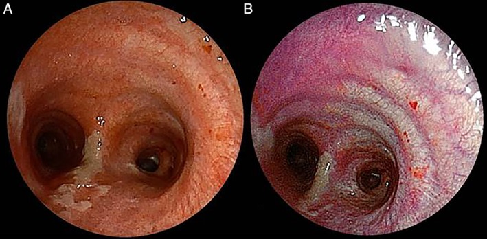
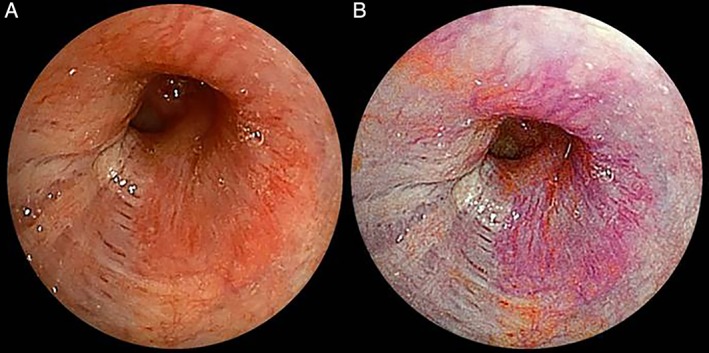
We report two cases of the comparison of diagnosis made with linked color imaging (LCI) and conventional white-light imaging (WLI) on the same patients. In case 1, a 75-year-old man in whom right upper lobectomy with mediastinal lymph node dissection was performed due to lung cancer had signs of bronchitis on postoperative day 8. The LCI demonstrated slight inflammatory changes that were not detectable with the conventional WLI on the tracheal wall. In case 2, in a 61-year-old woman who was diagnosed with adenoid cystic carcinoma, the bronchial wall was checked to confirm the extent of the tumour. The submucosal vascularity and tumour margin on the bronchial mucosa were better visible on LCI than on WLI. We could easily detect the mucosal inflammatory lesion and the malignant lesion with LCI in comparison with conventional WLI. Both mucosal inflammatory and malignant lesions were better visible with LCI in comparison to WLI.
Keywords: Bronchoscopy; inflammatory change; linked colour imaging; lung cancer.
Figures
References
-
- Fukuda H, Miura Y, Hayashi Y, et al. 2015. Linked color imaging technology facilitates early detection of flat gastric cancers. Clin. J. Gastroenterol. 8:385–389. - PubMed
-
- Satoh Y, Okumura S, Nakagawa K, et al. 2006. Postoperative ischemic change in bronchial stumps after primary lung cancer resection. Eur. J. Cardiothorac. Surg. 30:172–176. - PubMed
-
- Suzuki T, Hara T, Kitagawa Y, et al. 2017. Linked‐color imaging improves endoscopic visibility of colorectal nongranular flat lesions. Gastrointest. Endosc. 86:692–697. - PubMed
Publication types
LinkOut - more resources
Full Text Sources
