

Clinical and microstructural changes with different iontophoresis-assisted corneal cross-linking methods for keratoconus
- PMID: 30809476
- PMCID: PMC6376236
- DOI: 10.18240/ijo.2019.02.06
Clinical and microstructural changes with different iontophoresis-assisted corneal cross-linking methods for keratoconus
Abstract
Aim: To compare the clinical and microstructural changes induced by different transepithelial iontophoresis-assisted corneal cross-linking (I-CXL) methods for keratoconus.
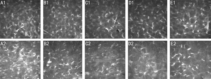
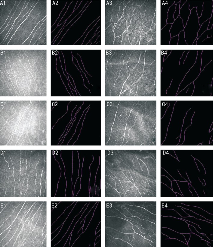
Methods: A total of 42 eyes of 42 patients with progressive keratoconus were divided into two groups. Group A received I-CXL for 5min, while group B received I-CXL for 10min. Visual acuity, optical coherence tomography (OCT), specular microscopy and confocal microscopy were evaluated preoperatively and at 1, 3, 6, and 12mo postoperatively.
Results: Twelve months after the operation, uncorrected visual acuity (UCVA) and corrected distance visual acuity (CDVA) were improved in both groups, with a better outcome in the I-CXL 10min group (P=0.025, 0.021, respectively). Kmax values decreased by 0.94±3.00 D in the I-CXL 10min group (P=0.033) but increased by 1.87±3.29 D in the I-CXL 5min group (P=0.012). OCT scans showed that the demarcation line was most visible and substantially deeper in the I-CXL 10min group. Confocal microscopy showed greater anterior stromal keratocyte decreases in the I-CXL 10min group than in the I-CXL 5min group at 3 and 6mo postoperatively (P<0.001); however, anterior stromal keratocytes and subbasal nerve density were not significantly different between the two groups at 12mo postoperatively.
Conclusion: I-CXL for 10min more effectively halts the progression of keratoconus than I-CXL for 5min after 12mo of follow-up. However, long-term studies are needed to evaluate the efficacy and safety of I-CXL.
Keywords: anterior stromal keratocyte; corneal cross-linking; iontophoresis; keratoconus; subbasal nerve density; transepithelial.
Figures
Similar articles
-
Optical coherence tomography and confocal microscopy following three different protocols of corneal collagen-crosslinking in keratoconus.Invest Ophthalmol Vis Sci. 2014 Oct 28;55(11):7601-9. doi: 10.1167/iovs.14-15662. Invest Ophthalmol Vis Sci. 2014. PMID: 25352122
-
[Transepithelial iontophoresis corneal collagen cross-linking for progressive keratoconus: one year results].Zhonghua Yan Ke Za Zhi. 2015 Sep;51(9):677-82. Zhonghua Yan Ke Za Zhi. 2015. PMID: 26693653 Chinese.
-
Conventional and Iontophoresis Corneal Cross-Linking for Keratoconus: Efficacy and Assessment by Optical Coherence Tomography and Confocal Microscopy.Cornea. 2017 Feb;36(2):153-162. doi: 10.1097/ICO.0000000000001062. Cornea. 2017. PMID: 28060061
-
Comparison of standard and accelerated corneal cross-linking for the treatment of keratoconus: a meta-analysis.Acta Ophthalmol. 2019 Feb;97(1):e22-e35. doi: 10.1111/aos.13814. Epub 2018 May 31. Acta Ophthalmol. 2019. PMID: 29855152 Review.
-
Efficacy and safety of transepithelial corneal collagen crosslinking surgery versus standard corneal collagen crosslinking surgery for keratoconus: a meta-analysis of randomized controlled trials.BMC Ophthalmol. 2017 Dec 28;17(1):262. doi: 10.1186/s12886-017-0657-2. BMC Ophthalmol. 2017. PMID: 29282020 Free PMC article. Review.
Cited by
-
Transepithelial corneal cross-linking assisted by two continuous cycles of iontophoresis for progressive keratoconus in adults: retrospective 5-year analysis.Graefes Arch Clin Exp Ophthalmol. 2021 Jan;259(1):239-246. doi: 10.1007/s00417-020-04861-y. Epub 2020 Jul 29. Graefes Arch Clin Exp Ophthalmol. 2021. PMID: 32725404 Free PMC article.
-
Efficacy, Safety, and Outcomes following Accelerated and Iontophoresis Corneal Crosslinking in Progressive Keratoconus.J Clin Med. 2023 Apr 18;12(8):2931. doi: 10.3390/jcm12082931. J Clin Med. 2023. PMID: 37109267 Free PMC article.
-
A Review of Keratoconus Cross-Linking Treatment Methods.J Clin Med. 2025 Mar 3;14(5):1702. doi: 10.3390/jcm14051702. J Clin Med. 2025. PMID: 40095727 Free PMC article. Review.
-
Safety and efficacy of repeated crosslinking assisted by transepithelial double-cycle iontophoresis in keratoconus progression after primary corneal crosslinking.Eye (Lond). 2021 Nov;35(11):3020-3027. doi: 10.1038/s41433-020-01365-1. Epub 2021 Jan 7. Eye (Lond). 2021. PMID: 33414527 Free PMC article.
-
A novel analysis of Scheimpflug total corneal refractive power following corneal cross-linking in mild to moderate keratoconus.Int J Ophthalmol. 2022 May 18;15(5):728-735. doi: 10.18240/ijo.2022.05.06. eCollection 2022. Int J Ophthalmol. 2022. PMID: 35601182 Free PMC article.
References
-
- Raiskup-Wolf F, Hoyer A, Spoerl E, Pillunat LE. Collagen crosslinking with riboflavin and ultraviolet-A light in keratoconus: long-term results. J Cataract Refract Surg. 2008;34(5):796–801. - PubMed
-
- Yildirim A, Cakir H, Kara N, Uslu H, Gurler B, Ozgurhan EB, Colak HN. Corneal collagen crosslinking for ectasia after laser in situ keratomileusis: long-term results. J Cataract Refract Surg. 2014;40(10):1591–1596. - PubMed
-
- Chan E, Snibson GR. Current status of corneal collagen cross-linking for keratoconus: a review. Clin Exp Optom. 2013;96(2):155–164. - PubMed
-
- Randleman JB, Khandelwal SS, Hafezi F. Corneal cross-linking. Surv Ophthalmol. 2015;60(6):509–523. - PubMed
LinkOut - more resources
Full Text Sources