Review
. 2018 Dec 13;5(1):52-67.
doi: 10.1016/j.ijwd.2018.08.011. eCollection 2019 Feb.

An approach to structural facial rejuvenation with fillers in women

Affiliations
Review

An approach to structural facial rejuvenation with fillers in women

R Fitzgerald et al. Int J Womens Dermatol. .

Abstract

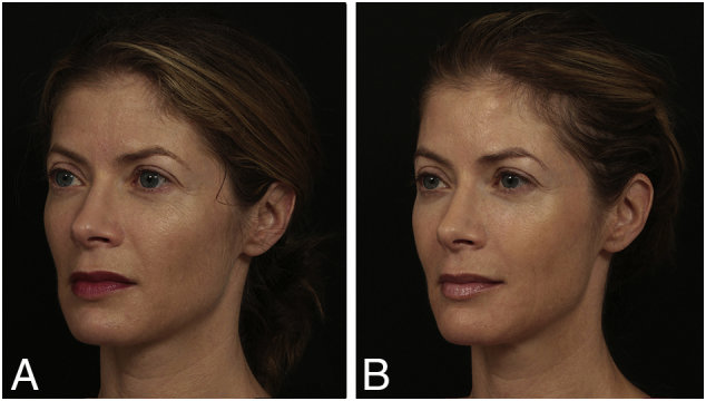
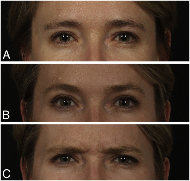
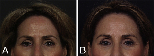
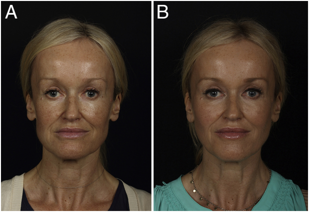
Newer understanding of volume loss as a critical component of facial aging and the integration of volume replacement into the surgical and nonsurgical therapeutic algorithm is arguably the most significant recent development in the field of facial rejuvenation. As all structural tissues play a role in the aging face, restoring youthful characteristics (or establishing them where they are congenitally absent) starts from the skeletal framework and builds progressively to the canvas of the face. The purpose of this article is to provide an introduction and brief summary of some of the current concepts concerning facial anatomy and the anatomy of facial aging, which serve as the basis for predictable and reproducible results with the use of injectable fillers. This article does not include the various types of fillers or techniques of filler injection, but covers how to decide where to use the filler and why, in different faces, as a result of the recognition and targeted correction of currently recognized specific anatomic deficiencies.

Keywords: Filler placement; anatomy of aging; facial analysis; facial fat compartments; facial volumization.


Figures

Fig. 1
Fig. 1
Timeline from 1800 to present, showing the date of the first description of important structures in the human face. DLCF, deep lateral cheek fat; DMCF, deep medial cheek fat; LOT, lateral orbital thickening; ROOF, retro-orbicularis oculi fat; SMAS, superficial musculoaponeurotic system; SOOF, suborbicularis oculi fat. Reproduced with permission from Cotofana et al. (2016).
Fig. 2
Fig. 2
Less volumized side of the face in patient with mild asymmetry simulates the effects of volume loss in all layers with aging. Reproduced with permission from Fitzgerald and Vleggaar (2009).
Fig. 3
Fig. 3
Facial proportions. (A) Vertical proportions are generally broken down into fifths based on the width of an eye. (B) Horizontal facial proportions are divided into thirds, measured from the hairline to the glabella, from the glabella to the subnasale, and from the subnasale to the menton. The lower third can be further broken down to the upper one-third, from the subnasale to the junction of the lips, and the lower two-thirds, from the junction of the lips to the menton. Reproduced with permission from Azizzadeh et al. (2007).
Fig. 4
Fig. 4
(Left) Average of facial surfaces of 116 female subjects, ages 20 to 30 years. (Right) Average of facial surfaces of 100 female subjects, ages 68 to 91 years (average, 76 years). Reproduced with permission from Lambros and Amos (2016). Three-dimensional facial averaging: Tool to understand facial aging. Image can be viewed animated online (http://links.lww.com/PRS/B922).
Fig. 5
Fig. 5
(A) The face is constructed of five basic layers. This five-layered construct is most evident in the scalp but exists in the rest of the face, with significant modification and compaction for functional adaptation. Layer 4 is the most significantly modified layer, with alternating facial soft tissue spaces and retaining ligaments. (B) To secure the superficial fascia to the facial skeleton, a system of retaining ligaments binds the dermis to the skeleton, and the components of this system pass through all layers. There are three morphological forms of retaining ligaments of the face. SMAS, superficial musculoaponeurotic system. Reproduced with permission from Mendelson and Wong (2013).
Fig. 6
Fig. 6
Individual variations in the onset and outcome of aging are commonplace. Many individuals age with relative parity in all structural tissue layers, but some are mostly a one tissue issue. (A) Young man with good bone structure and good skin, but extreme loss of fat. (B) Young woman with good skin and soft tissue volume, but congenital lack of craniofacial support. (C) Elderly woman who has good bone structure and ample soft tissue, but very elastotic skin. Photographs courtesy of Rebecca Fitzgerald, MD.
Fig. 7
Fig. 7
Schematic of the superficial and deep fat compartments. The superficial layer is composed of the nasolabial, medial cheek, middle cheek, and lateral temporal cheek compartments, as well as the three orbital compartments. The deep layer is composed of the suborbicularis oculi fat (medial SOOF and lateral SOOF) and the deep medial cheek fat (medial DMFC and lateral DLCF). A deep compartment termed Ristow’s space is located posterior to the most medial aspect of the deep medial cheek fat and lateral to the pyriform aperture. Reproduced with permission from Gierloff et al., 2012a, Gierloff et al., 2012b.
Fig. 8
Fig. 8
(A) Three-dimensional reconstruction of a contrast enhanced computed tomography (CT) scan. The superficial (subcutaneous) facial fat compartments have been injected with radiopaque dye. (B) Cadaveric dissection of the left side of a face after the same fat compartments have been injected with colored dye. Note that in this latter specimen, the tear trough, lateral orbital thickening, and area of the zygomatic arch did not stain. The dye did not migrate inferior to the nasolabial sulcus or medial to the labiomandibular sulcus. The superficial (subcutaneous) facial fat compartments shown are (1) superficial nasolabial, (2) medial cheek, (3) middle cheek, (4) lateral cheek, (5) superficial superior temporal, (6) superficial inferior temporal, and (7) jowl. The superficial central (8) and superficial lateral (9) forehead compartments are visualized in the CT image. Reproduced with permission from Schenck et al. (2018).
Fig. 9
Fig. 9
The upper third of the patient’s face is somewhat smaller and flatter than the lower two thirds, which looks more proportional and convex after treatment of the upper forehead at the hairline (using hyaluronic acid [HA]) with a 25-gauge cannula in the subgaleal space). The temples were also treated (HA placed on the bone with a 25-gauge needle). A very slight enhancement of the midface convexity was achieved by injecting HA into the prezygomatic space (25-gauge cannula), pyriform space (26-gauge needle after aspiration), and submentalis and labiomandibular fat compartments. On closer observation, the patient may have benefited from treatment in the superficial middle fat compartment (appreciated in the three quarter view) and the superficial lateral cheek compartment adjacent to the earlobe. Photographs courtesy of Rebecca Fitzgerald, MD.
Fig. 10
Fig. 10
(A) Patient (age 40s) was treated along her lateral upper lid and brow and her temples. (B) The after picture shows an anterior projection of the brows and lid after placement of product in the retro-orbicularis oculi fat (using hyaluronic acid and 25-gauge cannula), which gives the eye the youthful feature of a parallel brow, lid sulcus, and lash line. (C) Patient is shown frowning to demonstrate that this effect is not from a neuromodulator. Reproduced with permission from Azizzadeh et al. (2018).
Fig. 11
Fig. 11
(A) Patient lost temporal fat and superior preauricular fat area posterior to the hairline. Note that this affects the shape of her face, which is no longer 5-eyes across in the upper third. (B) This is restored with volume augmentation in this area. Reproduced with permission from Fitzgerald and Rubin (2014).
Fig. 12
Fig. 12
(A) Patient has some shadowing in her temples; however, the striking loss of lateral cheek fat is what gives the lateral area of her midface a concave appearance. (B) Treatment in this area restores an oval facial frame. The tragus is visible in an anterior view in a patient without lateral cheek fat and is less visible when this area is full. This defect is not uncommon in thin patients after face lifting. Hyaluronic acid was used with a 25-gauge cannula. Reproduced with permission from Azizzadeh et al. (2018).
Fig 13
Fig 13
(A) Deep midfacial fat compartments. The deep medial cheek fat is composed of a medial part and a lateral part (not shown). The medial part extends medially almost to the lateral incisor tooth. Augmentation of the deep medial cheek fat will consequently elevate and efface the nasolabial fold. The suborbicularis oculi fat is composed of a medial part and lateral part. With aging, inferior migration of these compartments occurs, as well as an inferior volume shift within the compartments, as seen on this computed tomography (CT) image. Reproduced from Gierloff et al., 2012a, Gierloff et al., 2012b. (B) Before and after hyaluronic acid filler was placed in the fat compartments shown in the CT image using a 25 gauge cannula. Improvement can be seen on the right side of the face in the three-quarter view. The patient was also treated along the lateral upper lid and brow as well as the temples, similar to the patient in Figure 10; however, neuromodulater was also used in the glabella and crow’s feet. Note the increased improvement when both filler and neuromodulater are used together. Reproduced with permission from Fitzgerald and Rubin (2014).
Fig. 14
Fig. 14
View of the right and left infraorbital region of layer 4 (deep to the OOM) in a female (A) and male (B) fresh-frozen specimen. (A) The PZ and lateral part of the deep cheek fat (DLCF) are circled. The hash symbol indicates the zygomaticus major muscle with its broadly based fibrous origin. This long attachment represents the lateral-inferior boundary of the DLCF. The facial vein is marked by the arrows and represents the medial-inferior boundary of the DLCF. The OOM is flipped toward the nose to expose the BB where this muscle attaches on the orbital rim. (B) Dye has been injected into the prezygomatic space (red and blue) and the lateral part of the DMCF (DLCF, green). The injection was performed with constant contact to the bone during application in all three locations. The red and blue areas correspond to the medial (MS) and lateral SOOF (LS). The hash symbol indicates zygomaticus major muscle with its broadly based fibrous origin; BB, bare bone area after sharp removal of the OOM from its attachment on the orbital rim. The asterisk marks the levator labii superioris alaeque nasi muscle. The arrows point to the facial vein. BB, Bare bone; DLCF, deep lateral cheek fat; DMCF, deep medial cheek fat; LS, lateral suborbicularis oculi fat; MS, medial suborbicularis oculi fat; OOM, orbicularis oculi muscle; PZ, prezygomatic space; SOOF, suborbicularis oculi fat. Reproduced with permission from Cotofana et al. (2015).
Fig. 15
Fig. 15
Clinical correlation of superficial and deep midfacial facial fat compartments. (A) In the first face pictured, deep midfacial fat is visible clinically. There is no visible undereye hollowing or nasolabial fold, and there is a convex contour to the midface. However, due to a congenital lipodystrophy, there is a striking lack of superficial fat. This is most obvious in the lateral temporal cheek compartment and upper periorbital area. However, on closer observation, the lack of superficial fat in the midface results in a good deal of shadowing in the lower lateral cheeks. When this face with ample deep midfacial fat is compared to the second face (B), the endurance exercise patient has only slight loss of the superficial fat, with only a small amount of shadowing in the temples and lateral cheeks, but a loss of fat in the deep midfacial compartments, which leads to a lack of midfacial projection and a very visible lid-cheek junction. Both patients are slightly asymmetric with the smaller side on their right. There is increased visibility of the zygomatic muscle on the smaller side of the first patient, and shape difference in the cheeks of the second patient with more deep midfacial fat on the left side, especially in the area of the lat SOOF and DLCF. Photographs courtesy of Rebecca Fitzgerald, MD.
Fig. 16
Fig. 16
(A) Before and (B) after camouflage of the nasojugal folds following volume augmentation of the four deep midfacial fat compartments (medial and lateral SOOF using hyaluronic acid and a 25-gauge cannula, and the deep medial cheek fat [DMCF] and deep lateral cheek fat [DLCF] using calcium hydroxylapatite and a 25-gauge cannula) is possible in this patient because she has strong midfacial bone structure, her orbital fat herniation does not exceed the volume loss in her deep midfacial fat, and her skin has good elasticity. On close observation, the difference in the volume of the DMCF and DLCF can be discerned between the larger (left) and smaller (right) sides of the face in the before picture. Photographs courtesy of Rebecca Fitzgerald, MD.
Fig. 17
Fig. 17
Just as there is suborbicularis fat around the eye, there is suborbicularis fat in the perioral region. Histologic examination confirms the macroscopic finding that the orbicularis insertion defines the wet-dry border of the lip. (A) Vertical sectioning of the upper lip reveals fat deep to the orbicularis oris muscle (arrow) and superficial to the buccal mucosa. (B) The clinical impression from this research is that the volume of deep lip fat contributes significantly to the appearance of the youthful lip. (C) Vertical sectioning of the lower lip shows deep submuscular fat (arrow). Of particular note, this specimen’s lower lip, which contained an ample amount of submuscular fat, showed anterior projection and eversion similar to that seen in a much younger individual. Reproduced with permission from Rohrich et al., 2008, Rohrich et al., 2009.
Fig. 18
Fig. 18
(A; top and bottom) After a previous injection, the patient’s lips appeared unnatural. (B; (top and bottom) Removing suboptimally placed filler in the vermillion border and body of the lip (with hyaluronidase) followed by placement of filler (using hyaluronic acid and a 25-gauge cannula) in a submuscular location in the mucosal and cutaneous portion of the patient’s upper and lower lip, as well as in the lateral and anterior chin, provide more support for the lips and a more natural-appearing result. Reproduced with permission from Fitzgerald and Rubin (2014).
Fig. 19
Fig. 19
(A) Volume-rendered three-dimensional spiral computed tomography (CT) image of the lower face, demonstrating the surface of the skin (above) and anterior part of the mandible with the labiomandibular fat compartments (LM) and the left inferior jowl fat (below). The yellow arrows indicate the position of the labiomandibular fold. The white arrow indicates the position of the mandibular retaining ligament. (B) Volume-rendered three-dimensional spiral CT image of the chin, demonstrating the submentalis fat. Note that this compartment does not lie immediately adjacent to the mentolabial sulcus. With permission from Gierloff et al., 2012a, Gierloff et al., 2012b.
Fig. 20
Fig. 20
Schematic illustration of the prezygomatic space (PZS), deep pyriform space, and important adjacent structures. Reproduced with permission from Surek et al. (2016).
Fig. 21
Fig. 21
(A) Layered dissection of the midface in an elderly male fresh cadaver after percutaneous injection of red dyed hyaluronic acid in the left hemiface. The orbicularis retaining ligament together with the periosteum of the orbital rim and orbital septum forms the arcus marginalis. In this dissection, the arcus marginalis was released to unveil the prezygomatic space. The superficial medial cheek fat is labeled for orientation. The prezygomatic capsule (PZC) is dyed with methylene blue. (B) The superficial medial cheek fat has been retracted to reveal the deep medial cheek fat and cavernous connection in the buccal recess. Reproduced with permission from Surek et al 2016.
Fig. 22
Fig. 22
(A) Patient with a shallow orbit, retrusive anterior maxilla, and inferior orbital rim. When the cornea has more anterior projection than the bony infraorbital rim, it is referred to as a negative vector, and a deep tear trough (TT) is often seen in these patients. The amount of filler required to correct the deep TT directly in these patients often results in an unnatural undereye contour and a Tyndall effect. A more natural-appearing correction can be achieved by providing volume support in the prezygomatic space and the pyriform aperature, as well as in the deep midfacial fat prior to treating the TT. (B) Patient after volumizing the prezygomatic space (hyaluronic acid administered with a 25 gauge cannula), which volumizes the inferiolateral rim and minimizes the amount of filler needed to fill the TT (which has not yet been filled). Photographs courtesy of Rebecca Fitzgerald, MD.
Fig. 23
Fig. 23
Deep pyriform space is cradled medially by the pyriform aperture and depressor septi nasi. The angular artery courses between the space and deep medial cheek fat compartment. Note that the artery is not directly on the periosteum, but rather superficial and lateral within the roof of the space. Postorbicularis oris fat is stained green. Reproduced with permission from Surek et al. (2016).
Fig. 24
Fig. 24
In this schematic, arrows indicate the areas of the craniofacial skeleton that are susceptible to resorption with aging. The size of the arrow correlates with the amount of resorption. Reproduced with permission from Mendelson and Wong (2012).
Fig. 25
Fig. 25
Relationship of the lip and nose affect overall facial balance. This schematic depicts the position of the lip in relation to the amount of pyriform support. Reproduced with permission from Yaremchuk 2007.
Fig. 26
Fig. 26
Clinical correlation of placement of product (here using poly-l-lactic acid/2 sessions) in the preperiosteal pyriform space, which pushes the base of the nose anteriorly and serves to evert the upper lip and increase the prominence of the cupid’s bow and philtral columns without direct augmentation of the lip. Product was also placed in the chin in the area of labiomandibular fat and deep submentalis fat, which serves to evert the lower lip. It is not uncommon to see the width of the lip shorten slightly with these injections. No product was used in the lips. Photographs courtesy of Rebecca Fitzgerald MD.
Fig. 27
Fig. 27
(A) Patient is shown before and (B) immediately after the first treatment, and (C) again 6 years later, maintained with annual treatments. Calcium hydroxylapatite administered with a 25-gauge cannula was used in the midface, and hyaluronic acid in the chin. All treatments were done with a 25-gauge cannula. A small chin implant was placed by a surgeon after the first treatment, but did not give the volume the patient desired. Since that time, hyaluronic acid was used after sterile scrub using care to avoid contact with the capsule of the implant. Change in the facial shape and perioral ratio is visible with this treatment. Photographs courtesy of Rebecca Fitzgerald, MD.
Fig. 28
Fig. 28
Practical use of these concepts in injectable treatment of the face can be illustrated using a number of patients of different ages and ethnic backgrounds. To examine a number of cases, as well as to compare and contrast different faces, these cases are presented in a “composite” format. This smaller format makes it easier to recognize facial shape and proportions and harmony or disharmony of the three thirds of the face, as well as to determine what is present or missing that may be moving the face away from the ideal shape and proportions illustrated in Figure 3. The photographs are lined up vertically to enable an easy comparison of the morphology of these faces both before and after treatment. In all cases, the facial shape is more ovalized and convex in the after pictures. Photographs courtesy of Rebecca Fitzgerald MD.
Supplementary material 12
Supplementary material 13
Supplementary material 14
Supplementary material 15
Supplementary material 16
Supplementary material 18
Supplementary material 19
Supplementary material 21
Supplementary material 22
Supplementary material 23
Supplementary material 26

References

    1. Avelar L.E., Cazerta C.E., Ave M.N., Shitara D.I. Dynamic changes of facial supporting cornerstones (pillars): Considerations in aesthetic approach. J Drugs Dermatol. 2018;17(4):466–470. - PubMed
    1. Azizzadeh B., Murphy M., Johnson C. Elsevier; 2007. Master techniques in facial rejuvenation.
    1. Azizzadeh B., Murphy M., Johnson C., Massry G., Fitzgerald R. Elsevier; London: 2018. Master techniques in facial rejuvenation.
    1. Bashour M. History and current concepts in the analysis of facial attractiveness. Plast Reconstr Surg. 2006;118:741–756. - PubMed
    1. Cotofana S., Schenck T.L., Trevidic P. Midface: clinical anatomy and regional approaches with injectable fillers. Plast Reconstr Surg. 2015;136.5S:219S–234S. - PubMed