

Review
doi: 10.1007/s00535-019-01552-2.
Epub 2019 Feb 26.
Recent advancement in EUS-guided fine needle sampling
Affiliations
- PMID: 30809717
- PMCID: PMC6470116
- DOI: 10.1007/s00535-019-01552-2
Item in Clipboard
Review
Recent advancement in EUS-guided fine needle sampling
J Gastroenterol.
2019 May.
Abstract
EUS-guided tissue acquisition technique plays an essential role for evaluation of gastrointestinal tumors. Several components affect the yield of EUS-guided tissue acquisition outcomes such as sampling techniques, use of ROSE (rapid onsite evaluation), training and experience, and needle designs. In this review we discuss advancement in EUS-guided fine needle sampling.
Keywords: Endoscopic ultrasound; Fine needle aspiration; Fine needle biopsy.
Figures
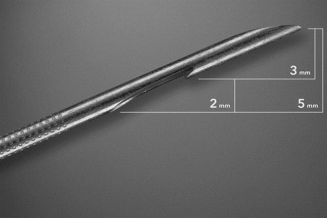
Image of EchoTip ProCore needle tip (22-gauge) showing the reverse bevel that promotes the procuring of core tissue sample from the target lesion (adapted from Dwyer et al. (2016), with permission)
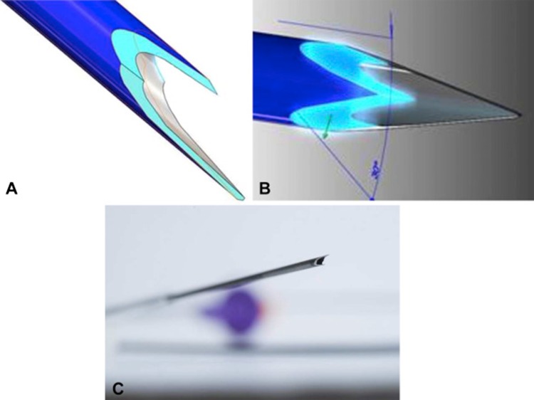
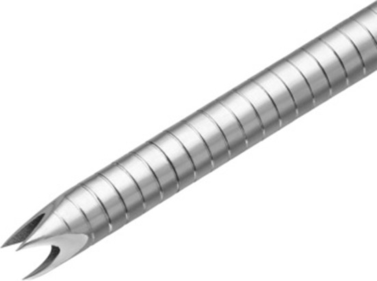
The 22-gauge tip of fork-tip needle with a second tip at opposite side of the lumen (adapted from Kandel et al. (2016), with permission)
The tip of 22-gauge Franseen needle design with a crown-shaped containing 3 symmetric planes (adapted from Bang et al. (2018), with permission)
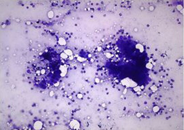
On-site cytological evaluation of pancreatic sample obtained by EUS–FNA supporting the diagnosis of pancreatic adenocarcinoma (adapted from Iglesias-Garcia et al. (2011), with permission)
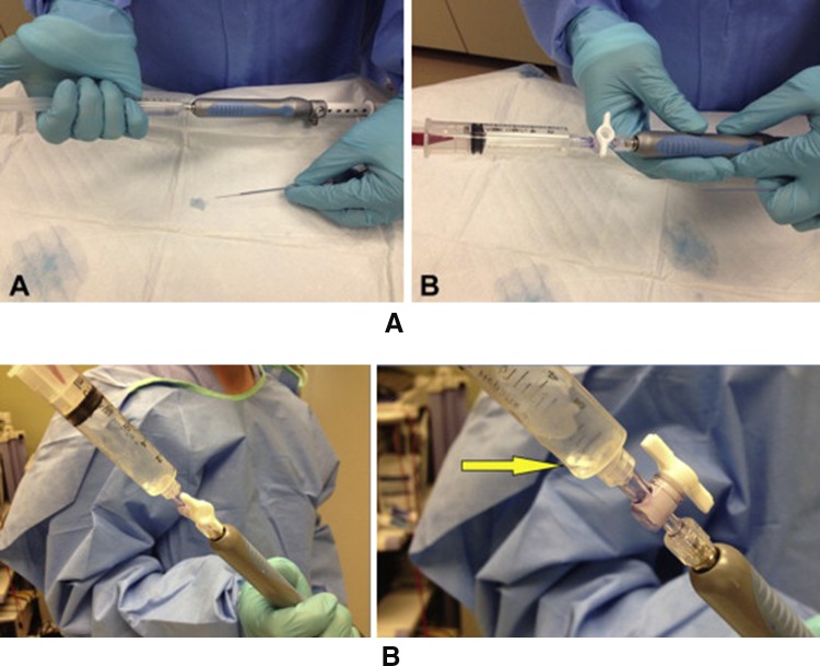
Demonstrating wet suction technique preparation with saline solution (a), loading suction syringe in locked position (b) and column of saline solution moving into the suction syringe as FNA is performed (adapted from Attam et al. (2015), with permission)
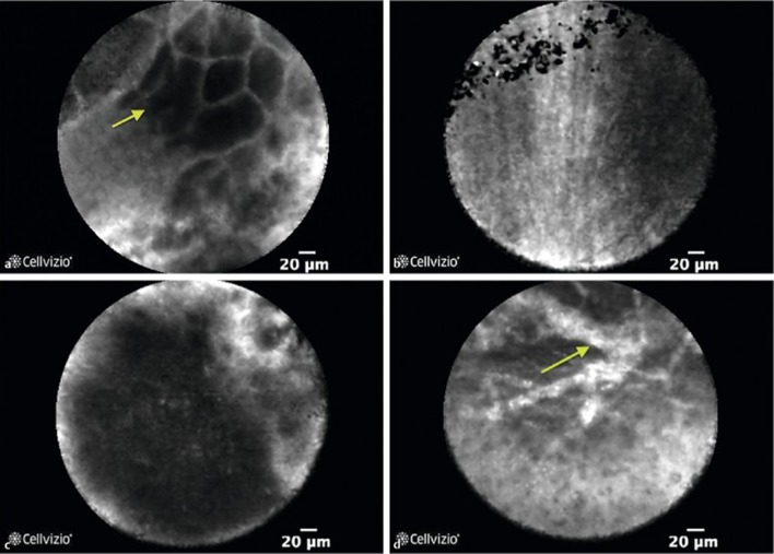
Needle-based confocal laser endomicroscopy (nCLE) images of benign and pancreatic ductal adenocarcinoma lesions. [Benign lesions: a showing normal acinar cells, b-showing fine white fibrous band representing a fibrotic tissue. Pancreatic ductal adenocarcinoma: c- showing dark aggregates > 40 μm, d- showing dilated vessels with fluorescein leakage] Adapted from Karstensen et al. (2018), with permission
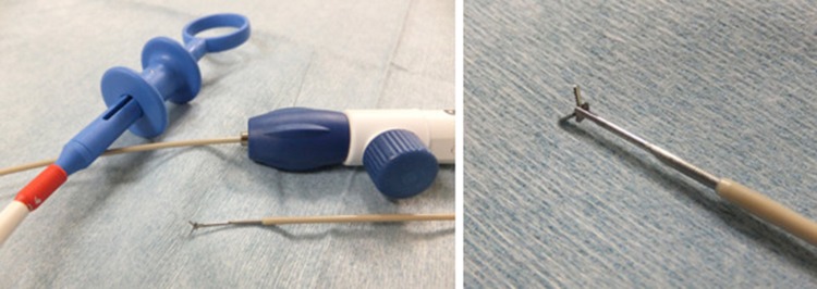
Small biopsy forceps through 19-gauge needle (adapted from Nakai et al. (2016), with permission)
Similar articles
-
Current status of newer generation endoscopic ultrasound core needles in the diagnostic evaluation of gastrointestinal lesions.J Am Soc Cytopathol. 2020 Sep-Oct;9(5):389-395. doi: 10.1016/j.jasc.2020.05.006. Epub 2020 Jun 5. J Am Soc Cytopathol. 2020. PMID: 32680792 Review.
-
Endoscopic ultrasound-guided fine-needle aspiration needles: which one and in what situation?Gastrointest Endosc Clin N Am. 2014 Jan;24(1):57-69. doi: 10.1016/j.giec.2013.08.008. Epub 2013 Sep 19. Gastrointest Endosc Clin N Am. 2014. PMID: 24215760 Review.
-
Techniques for endoscopic ultrasound-guided fine-needle biopsy.Gastrointest Endosc Clin N Am. 2014 Jan;24(1):83-107. doi: 10.1016/j.giec.2013.08.010. Gastrointest Endosc Clin N Am. 2014. PMID: 24215762 Review.
-
High clinical impact and diagnostic accuracy of EUS-guided biopsy sampling of subepithelial lesions: a prospective, comparative study.Surg Endosc. 2018 Mar;32(3):1304-1313. doi: 10.1007/s00464-017-5808-2. Epub 2017 Aug 15. Surg Endosc. 2018. PMID: 28812151 Free PMC article. Clinical Trial.
-
Efficacy of endoscopic-guided fine-needle aspiration in the diagnosis of gastrointestinal spindle cell tumors.Diagn Cytopathol. 2018 Aug;46(8):663-669. doi: 10.1002/dc.23976. Diagn Cytopathol. 2018. PMID: 31012545
Cited by
-
Minimally Invasive Sampling of Mediastinal Lesions.Life (Basel). 2024 Oct 11;14(10):1291. doi: 10.3390/life14101291. Life (Basel). 2024. PMID: 39459591 Free PMC article. Review.
-
Successful closure of a duodenal perforation caused by endoscopic ultrasound with an over-the-scope clip: a case report and literature review.J Int Med Res. 2023 Feb;51(2):3000605231154655. doi: 10.1177/03000605231154655. J Int Med Res. 2023. PMID: 36814402 Free PMC article. Review.
-
Diagnosis of gastric duplication cyst by positron emission tomography/computed tomography: A case report.World J Clin Cases. 2019 Nov 26;7(22):3866-3871. doi: 10.12998/wjcc.v7.i22.3866. World J Clin Cases. 2019. PMID: 31799316 Free PMC article.
-
Comparative diagnostic efficacy and safety of ultrasound-guided percutaneous transhepatic biopsy and endoscopic ultrasound-guided fine-needle aspiration biopsy for gallbladder tumors.Sci Rep. 2025 Apr 9;15(1):12155. doi: 10.1038/s41598-025-87847-2. Sci Rep. 2025. PMID: 40204763 Free PMC article.
-
Macroscopic on-site evaluation after EUS-guided fine needle biopsy may replace rapid on-site evaluation.Endosc Ultrasound. 2021 Mar-Apr;10(2):111-115. doi: 10.4103/EUS-D-20-00113. Endosc Ultrasound. 2021. PMID: 33885006 Free PMC article.
References
-
- Kandel P, Wallace MB. Optimizing endoscopic ultrasound guided tissue sampling of the pancreas. JOP J Pancreas. 2016;17:160–165.
-
- Early DS, Ben-Menachem T, Decker GA, et al. Aproppiate use of gastrointestinal endoscopy. Gastrointest Endosc. 2012;75:1127–1131. - PubMed
-
- Wani S, Muthusamy VR, Komanduri S. EUS-guided tissue acquisition: an evidence-based approach (with videos) Gastrointest Endosc. 2014;80(939–959):e937. - PubMed
-
- Fujii LL, Levy MJ. Pitfalls in EUS FNA. Gastrointest Endosc Clin N Am. 2014;24:125–142. - PubMed
Publication types
MeSH terms
LinkOut - more resources
Full Text Sources
