Review
. 2019 Feb;39(1):26-42.
doi: 10.1055/s-0038-1676806. Epub 2019 Jan 17.

Inflammation and Liver Cancer: Molecular Mechanisms and Therapeutic Targets

Affiliations
Review

Inflammation and Liver Cancer: Molecular Mechanisms and Therapeutic Targets

Yoon Mee Yang et al. Semin Liver Dis. 2019 Feb.

Abstract

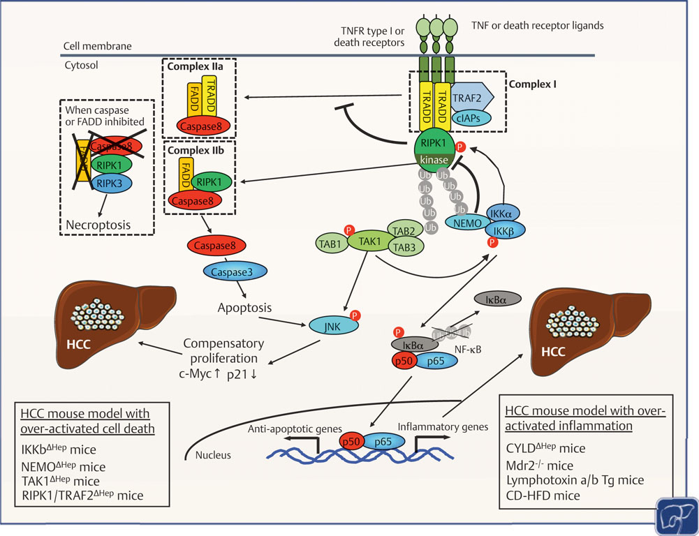
Hepatocellular carcinoma (HCC) is associated with chronic inflammation and fibrosis arising from different etiologies, including hepatitis B and C and alcoholic and nonalcoholic fatty liver diseases. The inflammatory cytokines tumor necrosis factor-α and interleukin-6 and their downstream targets nuclear factor kappa B (NF-κB), c-Jun N-terminal kinase (JNK), and signal transducer and activator of transcription 3 drive inflammation-associated HCC. Further, while adaptive immunity promotes immune surveillance to eradicate early HCC, adaptive immune cells, such as CD8+ T cells, Th17 cells, and B cells, can also stimulate HCC development. Thus, the role of the hepatic immune system in HCC development is a highly complex topic. This review highlights the role of cytokine signals, NF-κB, JNK, innate and adaptive immunity, and hepatic stellate cells in HCC and discusses whether these pathways could be therapeutic targets. The authors will also discuss cholangiocarcinoma and liver metastasis because biliary inflammation and tumor-associated stroma are essential for cholangiocarcinoma development and because primary tumor-derived inflammatory mediators promote the formation of a "premetastasis niche" in the liver.


Conflict of interest statement

Disclosure The authors report no conflicts of interest in this work.

Figures

Fig. 1
Fig. 1
Tumor necrosis factor (TNF) receptor, IkappaB kinase/nuclear factor kappa B (IKK/NF-κB), c-Jun N-terminal kinase (JNK), and apoptosis in hepatocellular carcinoma (HCC) development. In the development of inflammation-associated HCC, the IKK/NF-κB, JNK, and apoptosis pathways play both pro- and antitumorigenic roles. Activation of death receptors, including TNF receptor type I, leads to the formation of “complexI,” consisting of TNF receptor-associated death domain (TRADD), TNF receptor-associated factor 2 (TRAF2), and cellular inhibitor of apoptosis protein (cIAP). Polyubiquitination of receptor-interacting protein 1 (RIP1) recruits and activates the TAK1 and IKK complexes (IKKα/IKKβ/NF-kappa-B essential modulator [NEMO]). TAK1 activates the JNK mitogen-activated protein kinase (MAPK) pathway in a phosphorylation-dependent manner. TAK1 also phosphorylates and activates the IKK complex, leading to the phosphorylation, ubiquitination, and degradation of IκBα. This degradation results in the nuclear translocation and activation of NF-κB, comprising the p50 and p65 subunits. NF-κB promotes tumorigenesis by inducing inflammatory factors but prevents tumorigenesis by suppressing apoptotic pathways. NEMO regulates NF-κB activation and directly prevents the formation of the complex IIb (RIP kinase 1 [RIPK1], Fas-associated protein with death domain [FADD], caspase-8). RIPK1 kinase induces complex IIb-mediated hepatocyte apoptosis. NEMO and RIPK1 negatively regulate the formation of complex IIa (TRADD, FADD, caspase-8). Inhibition of caspase-8 and/or FADD leads to phosphorylation of RIP1 and RIP3 and induction of necroptosis. JNK is crucial for compensatory proliferation by increasing c-Myc and decreasing p21.
Fig. 2
Fig. 2
Interleukin (IL)-6 and the gender effect in hepatocellular carcinoma (HCC) development. In early HCC, IL-6 production is mediated by Toll-like receptor/MyD88 signaling in Kupffer cells and is negatively regulated by estrogen receptor α (ERα) signaling. NCOA5 is involved in ERα-mediated suppression of IL-6. IL-6 signaling promotes liver cancer cell growth by activating signal transducer and activator of transcription 3 (STAT3). IL-6 signaling also upregulates androgen receptor (AR) expression. AR inhibits the tumor suppressor p53 and enhances reactive oxygen species (ROS) production, promoting deoxyribonucleic acid (DNA) damage and mutation. In HCC progenitor cells, IL-6 is produced in an autocrine manner through upregulation of LIN28 and suppression of Let-7 and contributes to malignant transformation.
Fig. 3
Fig. 3
Anti- and protumorigenic roles of the immune cell network in hepatocellular carcinoma (HCC) development. Various immune cell networks have both anti- and protumorigenesis effects. Immune surveillance by natural killer (NK) cells, CD4+, CD8+ T cells, and B cells suppresses HCC development. However, the expansion of CD8+ T cells, immunoglobulin A (IgA)-producing plasma cells, Th17 cells, NKT cells, regulatory T cells, myeloid-derived suppressor cells (MDSCs), and M2 macrophages promotes HCC development. HSC senescence has been proposed to play both pro- and antitumorigenic roles by inducing the senescence-associated secretory phenotype (SASP) and by promoting M1 polarization, respectively.
Fig. 4
Fig. 4
miR-122 and miR-21 regulate inflammation-associated hepatocellular carcinoma (HCC). Tumor suppressor miR-122, the expression of which is reduced in HCC, is inhibited by tumor necrosis factor (TNF)-α or interleukin (IL)-6. Decreased miR-122 levels increase CCL2 levels, leading to the infiltration of inflammatory cells (e.g., CD11bhiGr1+ cells expressing CCR2) that produce TNFα and IL-6. Dysregulation of miR-122 induces BCL9 and many genes involved in proliferation and differentiation. miR-122 also negatively regulates the oncomiR miR-21. miR-21 is upregulated by IL-6 and CCL2. miR-21 targets PTEN, CAMSAP1, DDX1, MARCKSL1, and IL-12. miR-21 also targets programmed cell death 4 (PDCD4), creating a positive feedback loop. Dysregulation of miR-122 and miR-21 contributes to inflammation-associated HCC.

References

    1. Siegel RL, Miller KD, Jemal A. Cancer Statistics, 2018. CA Cancer J Clin 2018;68(01):7–30 - PubMed
    1. El-Serag HB, Rudolph KL. Hepatocellular carcinoma: epidemiology and molecular carcinogenesis. Gastroenterology 2007;132(07):2557–2576 - PubMed
    1. Massarweh NN, El-Serag HB. Epidemiology of hepatocellular carcinoma and intrahepatic cholangiocarcinoma. Cancer Contr 2017;24(03):1073274817729245 - PMC - PubMed
    1. Michelotti GA, Machado MV, Diehl AM. NAFLD, NASH and liver cancer. Nat Rev Gastroenterol Hepatol 2013;10(11):656–665 - PubMed
    1. Heymann F, Tacke F. Immunology in the liver–from homeostasis to disease. Nat Rev Gastroenterol Hepatol 2016;13(02):88–110 - PubMed

Publication types

MeSH terms

Supplementary concepts