Review
. 2019 Feb;11(1):15-24.
doi: 10.1111/os.12425. Epub 2019 Feb 27.

Bone Defects in Revision Total Knee Arthroplasty and Management

Affiliations
Review

Bone Defects in Revision Total Knee Arthroplasty and Management

Peng-Fei Lei et al. Orthop Surg. 2019 Feb.

Abstract

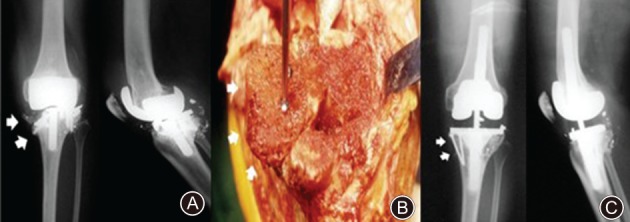
This article reviews the recent updates in revision of total knee arthroplasty (RTKA). We reviewed the recent articles on RTKA in databases including PubMed, Google Scholar, and SCOPUS. Total knee arthroplasty (TKA) involves the replacement of all three compartments of the knee in surgery of the knee joint to restore capacity and function. TKA is one of the most common and reliable surgical treatment options for the treatment of knee diseases. However, some patients require revision of TKA (RTKA) after primary TKA for various reasons, including mechanical wear, implant loosening or breakage, malalignment, infection, instability, periprosthetic fracture, and persistent stiffness. Unfortunately, the overall outcome of RTKA is not as satisfactory as for primary TKA due to the uncertainty regarding the actual success rate and the risk factors for failure. Cementation, modular metal augmentation, bone grafting, autologous bone grafting, allogenic bone grafting, impactation bone grafting, structural bone allografting, metaphyseal fixation, using porous titanium coated press fit metaphyseal sleeves and porous tantalum structural cones, and megaprostheses or customized prostheses are the currently available management options for RTKA. However, most of the management systems possess specific complications. Novel approaches should be developed to improve functional capacity, implant survival rates, and quality of life in a cost-efficient manner.

Keywords: Bone defects; Knee joint pain; Management; Revision of total knee arthroplasty.


Figures

Figure 1
Figure 1
The selection flow for included studies in this review. The first screening step is based on information provided by titles and abstracts, and 183 articles were excluded. The second screening step is to exclude the case report studies (n = 12) and the studies with incomplete data (n = 2). Finally, 60 articles were included.
Figure 2
Figure 2
Anderson Orthopaedic Research Institute classification of bone defects: (A) type I (intact metaphyseal bone with minor defects not compromising the stability of a revision component), (B) type IIA (damaged metaphyseal bone with defects in one femoral condyle or tibial plateau), (C) type IIB (more than one damaged metaphyseal bone), and (D) type III (deficient metaphyseal bone with bone loss compromising a major portion of the condyle or plateau). The latter defects are occasionally associated with collateral or patellar ligament detachment and usually require bone grafting or custom implants17.
Figure 3
Figure 3
Postoperative X‐ray for revision total knee arthroplasty using screw‐reinforced cement technique at the tibial side; with lateral translation at the femoral side.
Figure 4
Figure 4
(A, B) Postoperative X‐ray for revision total knee arthroplasty using modular metal augments for bone defects; (C, D) 25 months after revision total knee arthroplasty using modular metal augments for bone defects, there is no radiolucent line.
Figure 5
Figure 5
Preoperative anteroposterior (A) and lateral (B) radiographs of a right knee demonstrating an Anderson Orthopedic Research Institute type 3 tibial defect. Postoperative anteroposterior (C) and lateral (D) radiographs showing a revision with tibial allograft–implant composite. The tibial tubercle osteotomy is reattached to the allograft with a screw.
Figure 6
Figure 6
(A, B) Preoperative radiograph anteroposterior view and lateral view of a large tibial bone defects with instability. (C, D) Postoperative radiograph anteroposterior view and lateral view of short cemented stem with impacted bone graft for substantial tibial bone loss.
Figure 7
Figure 7
(A) Anteroposterior and lateral radiographs show a severe osteolysis in proximal tibia and distal femur with metal breakage (white arrow). (B) The femoral head allograft was stabilized with screws at the proximal tibia (white arrow). (C) The allograft remained intact with minimal resorption at 6 years after surgery (white arrow).
Figure 8
Figure 8
(A) Preoperative anteroposterior (AP) radiograph showing a Type IIA Anderson Orthopedic Research Institute tibial defect. (B) Postoperative AP radiograph (33 months) showing a cementless metaphyseal sleeve and stem construct.
Figure 9
Figure 9
(A) Optimized sizing and in situ fixation of femoral and tibial cone. (B) Use of a high‐speed burr to ensure an optimal fit of the femoral TM cone. (C) Pressfit impaction of a femoral metal cone in combination with a hinged implant.
Figure 10
Figure 10
(A) Preoperative radiograph showing complex bone defects. (B) Postoperative anteroposterior radiograph showing treatment with a megaprosthesis with a rotating hinge device severe bone defects.
Figure 11
Figure 11
The summary of current management of the bone defects in revision of total knee arthroplasty.

References

    1. Werner A, Jäger M, Schmitz H, Krauspe R. Joint preserving surgery for osteonecrosis and osteochondral defects after chemotherapy in childhood. Klin Padiatr, 2003, 215: 332–337. - PubMed
    1. Ji JH, Park SE, Song IS, Kang H, Ha JY, Jeong JJ. Complications of medial unicompartmental knee arthroplasty. Clin Orthop Surg, 2014, 6: 365–372. - PMC - PubMed
    1. Liddle AD, Judge A, Pandit H, Murray DW. Adverse outcomes after total and unicompartmental knee replacement in 101,330 matched patients: a study of data from the National Joint Registry for England and Wales. Lancet, 2014, 384: 1437–1445. - PubMed
    1. Rytter S, Egund N, Jensen LK, Bonde JP. Occupational kneeling and radiographic tibiofemoral and patellofemoral osteoarthritis. J Occup Med Toxicol, 2009, 4: 19. - PMC - PubMed
    1. Van Manen MD, Nace J, Mont MA. Management of primary knee osteoarthritis and indications for total knee arthroplasty for general practitioners. J Am Osteopath Assoc, 2012, 112: 709–715. - PubMed