

BRD4 inhibition attenuates inflammatory response in microglia and facilitates recovery after spinal cord injury in rats
- PMID: 30809946
- PMCID: PMC6484335
- DOI: 10.1111/jcmm.14196
BRD4 inhibition attenuates inflammatory response in microglia and facilitates recovery after spinal cord injury in rats
Abstract
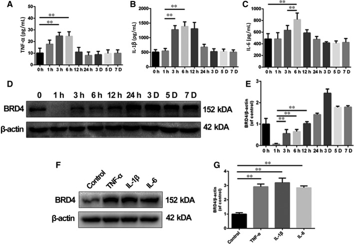
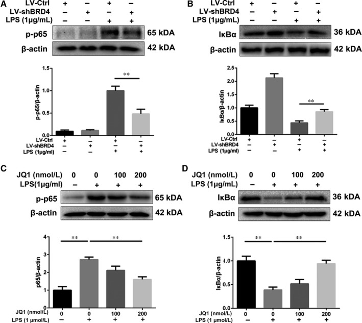
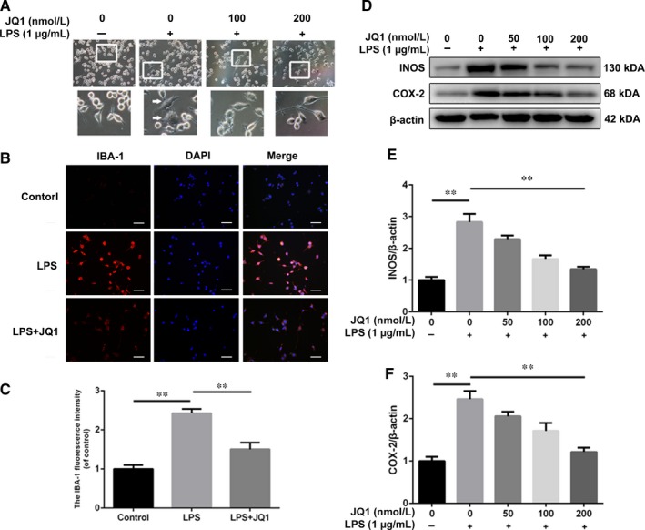
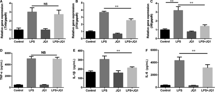
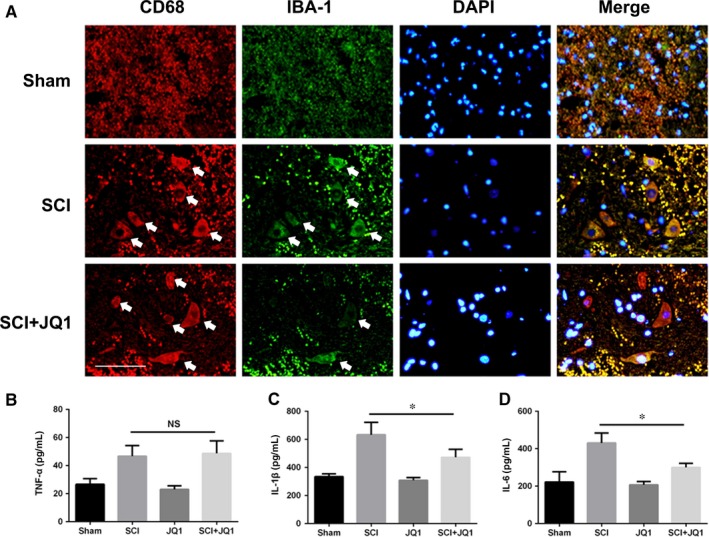
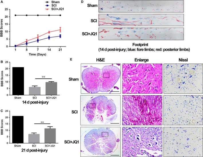
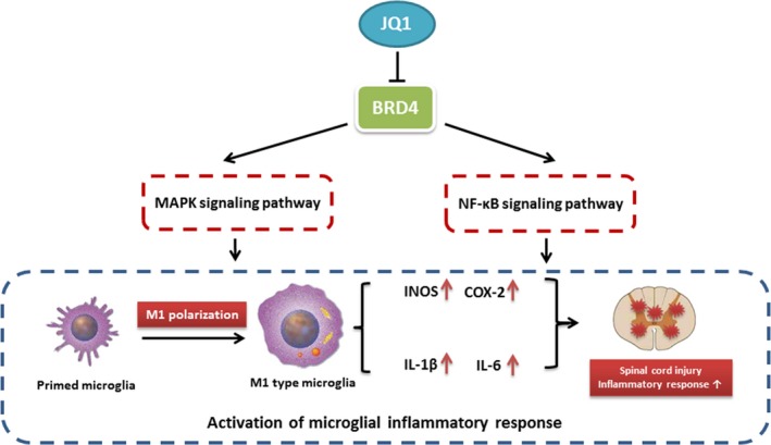
The pathophysiology of spinal cord injury (SCI) involves primary injury and secondary injury. For the irreversibility of primary injury, therapies of SCI mainly focus on secondary injury, whereas inflammation is considered to be a major target for secondary injury; however the regulation of inflammation in SCI is unclear and targeted therapies are still lacking. In this study, we found that the expression of BRD4 was correlated with pro-inflammatory cytokines after SCI in rats; in vitro study in microglia showed that BRD4 inhibition either by lentivirus or JQ1 may both suppress the MAPK and NF-κB signalling pathways, which are the two major signalling pathways involved in inflammatory response in microglia. BRD4 inhibition by JQ1 not only blocked microglial M1 polarization, but also repressed the level of pro-inflammatory cytokines in microglia in vitro and in vivo. Furthermore, BRD4 inhibition by JQ1 can improve functional recovery and structural disorder as well as reduce neuron loss in SCI rats. Overall, this study illustrates that microglial BRD4 level is increased after SCI and BRD4 inhibition is able to suppress M1 polarization and pro-inflammatory cytokine production in microglia which ultimately promotes functional recovery after SCI.
Keywords: BRD4; JQ1; inflammation; microglia; spinal cord injury.
© 2019 The Authors. Journal of Cellular and Molecular Medicine published by John Wiley & Sons Ltd and Foundation for Cellular and Molecular Medicine.
Conflict of interest statement
The authors declare no conflicts of interest.
Figures

Similar articles
-
Bromodomain and extraterminal domain-containing protein inhibition attenuates acute inflammation after spinal cord injury.Exp Neurol. 2018 Nov;309:181-192. doi: 10.1016/j.expneurol.2018.08.005. Epub 2018 Aug 19. Exp Neurol. 2018. PMID: 30134146
-
Salidroside attenuates neuroinflammation and improves functional recovery after spinal cord injury through microglia polarization regulation.J Cell Mol Med. 2018 Feb;22(2):1148-1166. doi: 10.1111/jcmm.13368. Epub 2017 Nov 17. J Cell Mol Med. 2018. PMID: 29148269 Free PMC article.
-
BRD4 suppression alleviates cerebral ischemia-induced brain injury by blocking glial activation via the inhibition of inflammatory response and pyroptosis.Biochem Biophys Res Commun. 2019 Nov 12;519(3):481-488. doi: 10.1016/j.bbrc.2019.07.097. Epub 2019 Sep 14. Biochem Biophys Res Commun. 2019. PMID: 31530390
-
JQ1: a novel potential therapeutic target.Pharmazie. 2018 Sep 1;73(9):491-493. doi: 10.1691/ph.2018.8480. Pharmazie. 2018. PMID: 30223929 Review.
-
The NF-κB Pathway: a Focus on Inflammatory Responses in Spinal Cord Injury.Mol Neurobiol. 2023 Sep;60(9):5292-5308. doi: 10.1007/s12035-023-03411-x. Epub 2023 Jun 7. Mol Neurobiol. 2023. PMID: 37286724 Review.
Cited by
-
BET bromodomains as novel epigenetic targets for brain health and disease.Neuropharmacology. 2020 Dec 15;181:108306. doi: 10.1016/j.neuropharm.2020.108306. Epub 2020 Sep 15. Neuropharmacology. 2020. PMID: 32946883 Free PMC article. Review.
-
Salvianic acid A alleviates chronic alcoholic liver disease by inhibiting HMGB1 translocation via down-regulating BRD4.J Cell Mol Med. 2020 Aug;24(15):8518-8531. doi: 10.1111/jcmm.15473. Epub 2020 Jun 29. J Cell Mol Med. 2020. PMID: 32596881 Free PMC article.
-
Advances in the research of the role of macrophage/microglia polarization-mediated inflammatory response in spinal cord injury.Front Immunol. 2022 Dec 1;13:1014013. doi: 10.3389/fimmu.2022.1014013. eCollection 2022. Front Immunol. 2022. PMID: 36532022 Free PMC article.
-
The BET inhibitor attenuates the inflammatory response and cell migration in human microglial HMC3 cell line.Sci Rep. 2021 Apr 23;11(1):8828. doi: 10.1038/s41598-021-87828-1. Sci Rep. 2021. PMID: 33893325 Free PMC article.
-
miR-29c-3p downregulation accelerates spinal cord injury progression by targeting BRD4.J Orthop Surg Res. 2025 Jul 25;20(1):703. doi: 10.1186/s13018-025-06108-0. J Orthop Surg Res. 2025. PMID: 40713817 Free PMC article.
References
-
- Ahuja CS, Wilson JR, Nori S, et al. Traumatic spinal cord injury. Nat Rev Dis Primers. 2017;3:17018. - PubMed
-
- Blight AR. Delayed demyelination and macrophage invasion: a candidate for secondary cell damage in spinal cord injury. Cent Nerv Syst Trauma. 1985;2:299‐315. - PubMed
-
- Tator CH. Update on the pathophysiology and pathology of acute spinal cord injury. Brain Pathol. 1995;5:407‐413. - PubMed
Publication types
MeSH terms
Substances
LinkOut - more resources
Full Text Sources
Medical
