

Hepatic splenosis: Rare yet important - A case report and literature review
- PMID: 30810057
- PMCID: PMC6460629
- DOI: 10.1177/0300060519828901
Hepatic splenosis: Rare yet important - A case report and literature review
Abstract
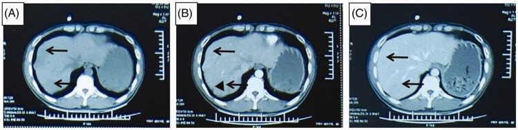
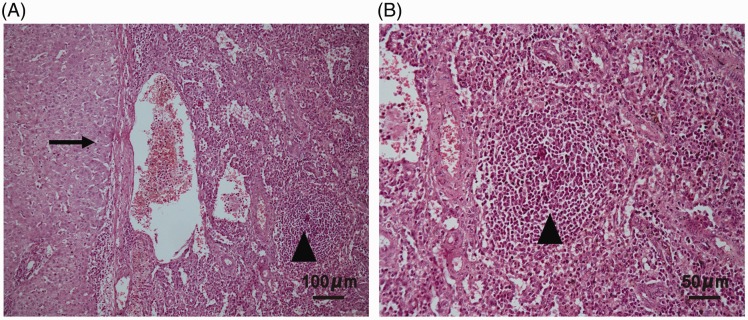
Hepatic splenosis is an uncommon condition that occurs following traumatic splenic rupture or splenectomy. The case of a 41-year-old male patient with multiple isolated liver masses indistinguishable from primary and metastatic liver tumours is reported. Following laparotomy, the liver lesions were resected and histopathology confirmed a diagnosis of hepatic splenosis. At an 18-month follow-up examination, no abnormalities in routine blood test, liver function, and liver computed tomography (CT) scanning were observed. After review of the literature, the following diagnostic criteria for hepatic splenosis are proposed: (1) a history of splenic trauma or splenectomy; (2) lesion(s) with a surrounding rim, particularly near the liver capsule identified by CT scanning; (3) findings on superparamagnetic iron oxide-enhanced magnetic resonance imaging or technetium-99m heat-damaged red cell scanning; and (4) histopathological findings (needle biopsy or surgical pathology). The following diagnostic process is also proposed: suspect diagnosis when criteria 1 and 2 are met; make diagnosis when criterion 3 is met; confirm diagnosis when criterion 4 is met. Laparotomy is recommended for either diagnosis or treatment when invasive procedures are necessary.
Keywords: Hepatic splenosis; diagnosis; treatment.
Figures
References
-
- Livingston CD, Levine BA, Lecklitner ML, et al. Incidence and function of residual splenic tissue following splenectomy for trauma in adults. Arch Surg 1983; 118: 617–620. - PubMed
-
- Ksiadzyna D, Peña AS. Abdominal splenosis. Rev Esp Enferm Dig 2011; 103: 421–426. - PubMed
-
- Hong SH, Kim JE, Cho ML, et al. Comparison of the Child-Turcotte-Pugh classification and the model for end-stage liver disease score as predictors of the severity of the systemic inflammatory response in patients undergoing living-donor liver transplantation. J Korean Med Sci 2011; 26: 1333–1338. - PMC - PubMed
-
- Albrecht H. A case of very numerous, over the whole peritoneum, scattered accessory spleens. Beitr z Path Anat 1896; 20: 513–527 [In German].
Publication types
MeSH terms
LinkOut - more resources
Full Text Sources
Medical
Miscellaneous
