. 2019 May 1;316(5):L843-L868.
doi: 10.1152/ajplung.00416.2018. Epub 2019 Feb 27.

Regulation of IL-17A and implications for TGF-β1 comodulation of airway smooth muscle remodeling in severe asthma

Affiliations

Regulation of IL-17A and implications for TGF-β1 comodulation of airway smooth muscle remodeling in severe asthma

Jon M Evasovic et al. Am J Physiol Lung Cell Mol Physiol. .

Abstract

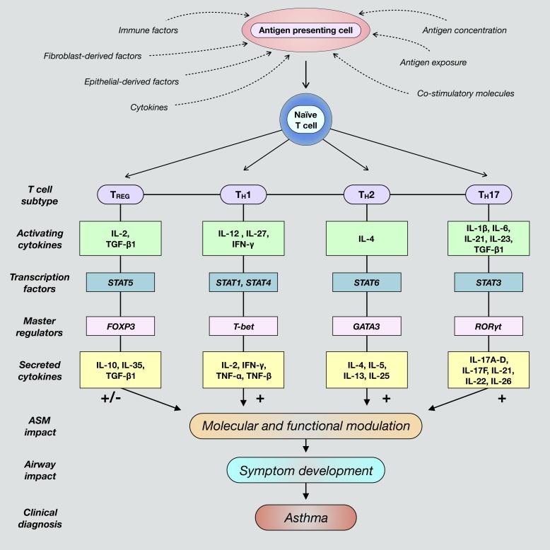
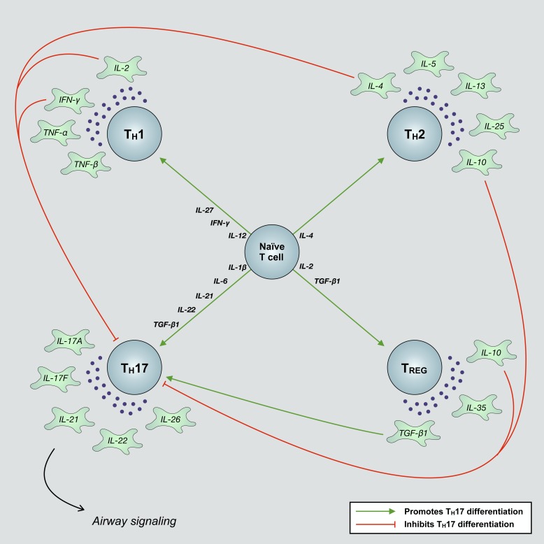
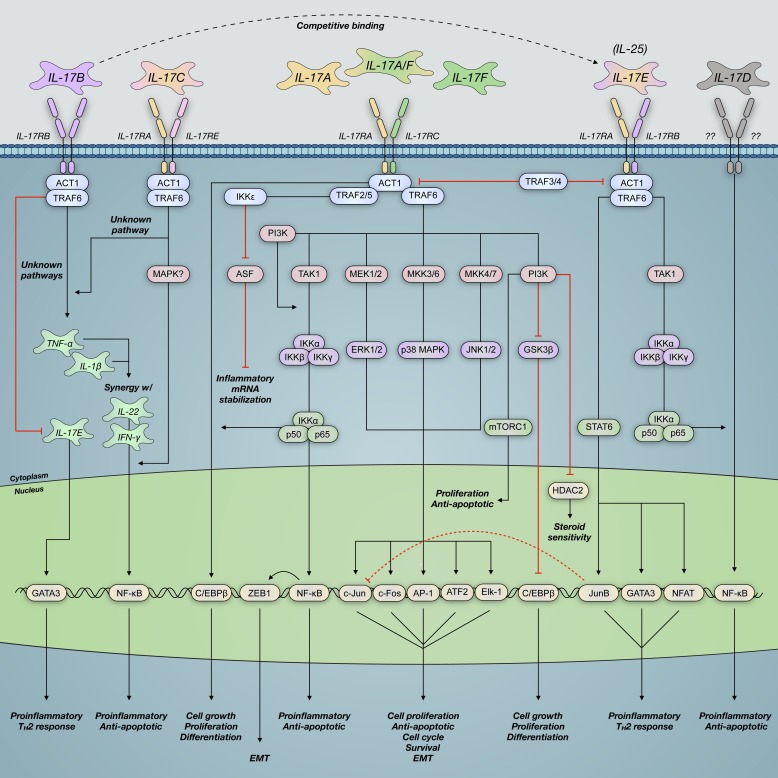
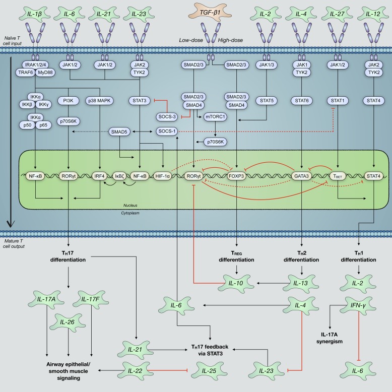
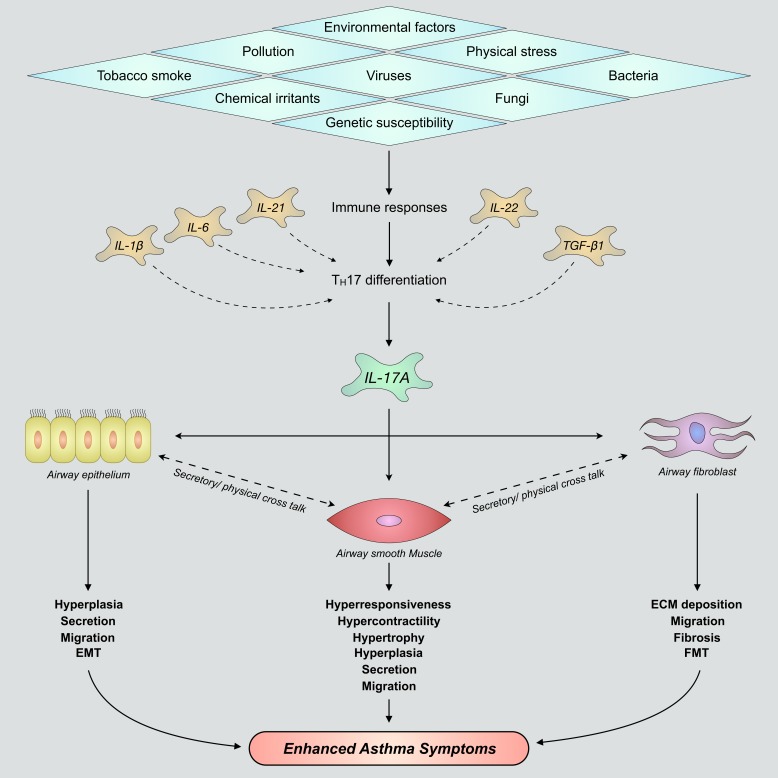
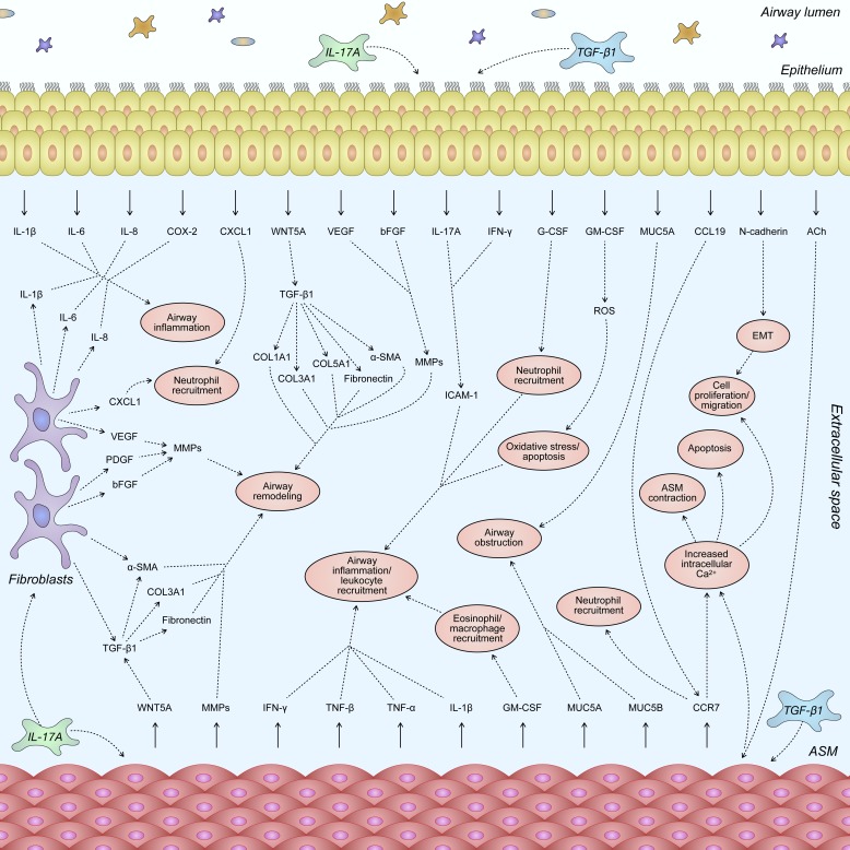
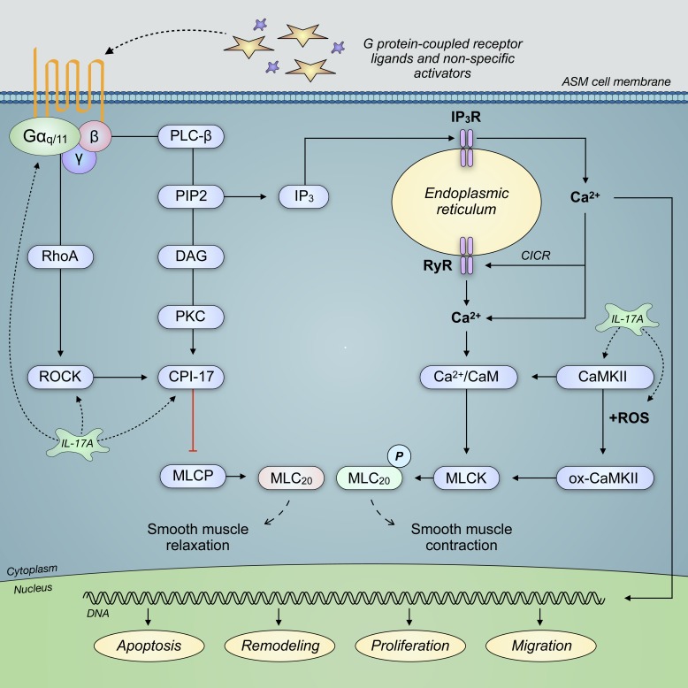
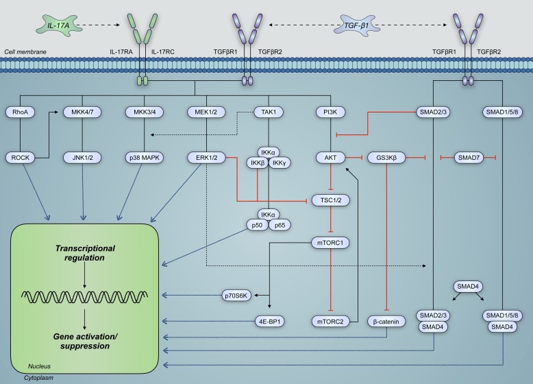
Severe asthma develops as a result of heightened, persistent symptoms that generally coincide with pronounced neutrophilic airway inflammation. In individuals with severe asthma, symptoms are poorly controlled by high-dose inhaled glucocorticoids and often lead to elevated morbidity and mortality rates that underscore the necessity for novel drug target identification that overcomes limitations in disease management. Many incidences of severe asthma are mechanistically associated with T helper 17 (TH17) cell-derived cytokines and immune factors that mediate neutrophilic influx to the airways. TH17-secreted interleukin-17A (IL-17A) is an independent risk factor for severe asthma that impacts airway smooth muscle (ASM) remodeling. TH17-derived cytokines and diverse immune mediators further interact with structural cells of the airway to induce pathophysiological processes that impact ASM functionality. Transforming growth factor-β1 (TGF-β1) is a pivotal mediator involved in airway remodeling that correlates with enhanced TH17 activity in individuals with severe asthma and is essential to TH17 differentiation and IL-17A production. IL-17A can also reciprocally enhance activation of TGF-β1 signaling pathways, whereas combined TH1/TH17 or TH2/TH17 immune responses may additively impact asthma severity. This review seeks to provide a comprehensive summary of cytokine-driven T cell fate determination and TH17-mediated airway inflammation. It will further review the evidence demonstrating the extent to which IL-17A interacts with various immune factors, specifically TGF-β1, to contribute to ASM remodeling and altered function in TH17-driven endotypes of severe asthma.

Keywords: IL-17A; TGF-β1; airway remodeling; airway smooth muscle; asthma.


Conflict of interest statement

No conflicts of interest, financial or otherwise, are declared by the authors.

Figures

Fig. 1.
Fig. 1.
Differentiation of naïve cluster of differentiation 4-positive (CD4+) T cells toward polarized, individual effector T cell fates is determined by the presence of unique activating factors and molecules present within a given inflammatory airway microenvironment. These commonly include various factors secreted by epithelial cells and fibroblasts, such as cytokines and chemokines. Additionally, T cell differentiation is promoted when antigen-presenting cells are exposed to foreign antigens and subsequently interact with naïve T cells. Classical T cell subtypes involved in airway immunity include regulatory T (TREG), T helper 1 (TH1), TH2, and TH17 cells. Mature T cells are characterized by the expression and activation of subtype-specific transcription factors and master regulators that ultimately induce secretion of unique cytokines contingent upon differentiation fate. These cytokines activate cognate receptors expressed by structural airway cells that direct immune responses and contribute to dysregulated airway smooth muscle (ASM) function and asthma pathogenesis. FOXP3, forkhead box protein P3; GATA3, trans-acting T cell-specific transcription factor GATA-3; RORγt, retinoic acid receptor (RAR)-related orphan receptor-γ, thymus; T-bet, T cell-specific T-box transcription factor T-bet; TGF-β1, transforming growth factor-β1.
Fig. 2.
Fig. 2.
Various immune and structural airway cells secrete inflammatory factors that promote the differentiation of naïve T cells into mature effector subtypes. These T cells further secrete subtype-dependent cytokines that bear the ability to promote and/or suppress the differentiation of disparate T cell subtypes. T helper 1 (TH1)- and TH2-derived IL-2, IL-4, IL-10, and IFN-γ inhibit the differentiation of naïve T cells into mature TH17 effector cells. Regulatory T (TREG) cells, depending on the concentration of their secreted cytokines, transforming growth factor-β1 (TGF-β1) and IL-10, can either promote or inhibit polarization toward a TH17 fate. Observations of such mechanisms, which ultimately alter airway function, underscore the complex immune signaling networks that regulate T cell differentiation and may in part explain the acquisition of mixed T cell endotypes in distinct individuals with asthma.
Fig. 3.
Fig. 3.
IL-17 family cytokines signal through heterodimeric IL-17 surface receptor subunits. This results in NF-κB activator (ACT)/TNF receptor-associated factor (TRAF) recruitment and activation of downstream effectors that impact gene transcription airway functionality. IL-17A binds its cognate IL-17 receptor A and C (IL-17RA/C) complex and activates mitogen-associated protein kinase (MAPK), phosphatidylinositol 4,5-bisphosphate 3-kinase (PI3K), and nuclear factor-κ-light chain enhancer of activated B cells (NF-κB) signaling pathways. These pathways contribute to altered airway smooth muscle function, airway remodeling, and development of symptoms consistent with severe asthma. Although other IL-17 cytokines signal via similar pathways, their mechanisms and functions within the asthmatic airway remain poorly characterized compared with those of IL-17A. AP-1, transcription factor AP-1; ASF, arginine- and serine-rich splicing factor; ATF2, cyclic AMP-dependent transcription factor ATF-2; C/EBPβ, CCAAT/enhancer-binding protein-β; Elk-1, E26 transformation-specific (ETS) domain-containing protein Elk-1; EMT, epithelial-mesenchymal transition; GATA3, trans-acting T cell-specific transcription factor GATA-3; HDAC2, histone deacetylase 2; mTORC1, mammalian target of rapamycin complex 1; NFAT, nuclear factor of activated T cells; TAK1, transforming growth factor-β (TGF-β)-activated kinase 1; TH2, T helper 2; w/, with; ZEB1, zinc finger E-box-binding homeobox 1.
Fig. 4.
Fig. 4.
T helper 17 (TH17) cell differentiation relies on the coordination of several well-characterized cytokines and their activation of cognate receptors on the surface of naïve T cells. IL-6 and transforming growth factor-β1 (TGF-β1) are the most prominent drivers of TH17 polarization via canonical signal transducer mothers against decapentaplegic homolog (SMAD) and signal transducer and activator of transcription 3 (STAT3) pathways. This enhances activation of TH17-necessary transcription factor retinoic acid receptor (RAR)-related orphan receptor-γ, thymus (RORγt). Other cytokines, including IL-1β, IL-21, and IL-23, can further drive TH17 differentiation and, moreover, promote their sustained maintenance. Regulatory T (TREG)-, TH1-, and TH2-associated cytokines typically activate signaling pathways that repress RORγt activation and inhibit TH17 differentiation. However, depending on the precise composition of cytokines exposed to naïve T cell surface receptors and interactions between intracellular signaling pathways, there is evidence of increased TH17 differentiation by these cytokines. FOXP3, forkhead box protein P3; HIF-1α, hypoxia-inducible factor 1α; IRAK, IL-1 receptor-associated kinase; IRF-4, interferon regulatory factor 4; mTORC1, mammalian target of rapamycin complex 1; MyD88; myeloid differentiation primary response protein MyD88; p70S6K, ribosomal protein S6 kinase β-1; PI3K, phosphatidylinositol 4,5-bisphosphate 3-kinase; SOCS, suppressor of cytokine signaling; T-bet, T cell-specific T-box transcription factor T-bet; TRAF6, TNF receptor-associated factor 6; TYK2, nonreceptor tyrosine-protein kinase TYK2.
Fig. 5.
Fig. 5.
Nonspecific inflammatory stimuli induce immune responses that promote T helper 17 (TH17) cell differentiation and production/secretion of IL-17A within immune-infiltrated airways. IL-17A variably acts upon airway epithelium, airway smooth muscle, and lung fibroblast cells to promote elevated airway remodeling and development of severe asthma. Although each of these structural cells imparts a unique effect on airway remodeling following stimulation with IL-17A, there is significant cellular cross talk between tissues. This bears potential to further enhance TH17-mediated effects surrounding airway smooth muscle hyperplasia and hypertrophy, goblet cell mucus secretions, epithelial-mesenchymal transition (EMT), extracellular matrix (ECM) deposition, fibroblast-to-myofibroblast transition (FMT), and airway hyperresponsiveness. TGF-β1, transforming growth factor-β1.
Fig. 6.
Fig. 6.
IL-17A and/or transforming growth factor-β1 (TGF-β1) signaling induce secretion of an array of inflammatory factors from airway epithelial, smooth muscle, and fibroblast cells. Moreover, several of these factors promote enhanced airway remodeling and phenotypic switching between structural airway cells. Dynamic cellular cross talk between cells within the airway architecture elevates expression of remodeling proteins beyond that seen within isolated cells following IL-17A and/or TGF-β1 treatment. In individuals with severe asthma, these interactions are suggested to be responsible for increased cellular migration, reduced epithelial integrity, thickened airway smooth muscle (ASM) layers, and a marked degree of airway obstruction and reduced airflow. bFGF, basic fibroblast growth factor; CCL19, C-C motif chemokine 19; CCR7, C-C chemokine receptor 7; COL, collagen; COX-2, cyclooxygenase-2; CXCL1, C-X-C motif chemokine ligand 1; EMT, epithelial-mesenchymal transition; G-CSF, granulocyte colony-stimulating factor; GM-CSF, granulocyte-macrophage colony-stimulating factor; MMPs, matrix metalloproteinases; MUC, mucin; ROS, reactive oxygen species; α-SMA, α-smooth muscle actin; WNT5A, protein Wnt-5a.
Fig. 7.
Fig. 7.
IL-17A signaling is correlated with enhanced expression and activation of Ras homolog gene family, member A (RhoA), Rho-associated protein kinase (ROCK), C-kinase-activated protein phosphatase-1 inhibitor (CPI-17), and Ca2+/calmodulin-dependent protein kinase II (CaMKII). These G protein-coupled receptor effectors regulate the phosphorylation (P) of myosin light chain, which modulates smooth muscle contraction. Enhanced contractile force of airway smooth muscle (ASM) cells is known to lead to airway obstruction through a reduced airway lumen diameter. Moreover, oxidized CaMKII (ox-CaMKII) is a product of CaMKII interaction with reactive oxygen species (ROS), which may promote airway reactivity. CICR, calcium-induced calcium release; DAG, diacylglycerol; IP3, inositol (1,4,5)-trisphosphate; IP3R, IP3 receptor; MLC20, myosin regulatory light chain 20 kDa; MLCK, myosin light chain kinase; MLCP, myosin light chain phosphatase; PIP2, phosphatidylinositol 4,5-bisphosphate; RyR, ryanodine receptor.
Fig. 8.
Fig. 8.
IL-17A and transforming growth factor-β1 (TGF-β1) share significant cross talk in the regulation and activation of signaling pathways pertinent to structural airway cell function. These signaling pathways, which are intricately connected, can modulate the activity of one another such that overall transcriptional regulation is altered. Moreover, TGF-β1 signaling activates signal transducer mothers against decapentaplegic homolog (SMAD) pathways, which can interact with IL-17A-activated pathways to modulate gene expression. Such pathways include those of Ras homolog gene family, member A (RhoA), mitogen-activate protein kinases (MAPKs), and phosphatidylinositol 4,5-bisphosphate 3-kinase (PI3K). 4E-BP1, eukaryotic translation initiation factor 4E-binding protein-1; GS3Kβ, glycogen synthase kinase-3β; IL-17RA and IL-17RC, IL-17 receptors A and C, respectively; mTORC, mammalian target of rapamycin complex; p70S6K, ribosomal protein S6 kinase β-1; ROCK, Rho-associated protein kinase; TAK1, TGF-β-activated kinase 1; TGFBR, TGF-β receptor; TSC, tuberous sclerosis protein.

Similar articles

Cited by

References

    1. Agache I, Ciobanu C, Agache C, Anghel M. Increased serum IL-17 is an independent risk factor for severe asthma. Respir Med 104: 1131–1137, 2010. doi:10.1016/j.rmed.2010.02.018. - DOI - PubMed
    1. Al-Alawi M, Hassan T, Chotirmall SH. Transforming growth factor β and severe asthma: a perfect storm. Respir Med 108: 1409–1423, 2014. doi:10.1016/j.rmed.2014.08.008. - DOI - PubMed
    1. Alrashdan YA, Alkhouri H, Chen E, Lalor DJ, Poniris M, Henness S, Brightling CE, Burgess JK, Armour CL, Ammit AJ, Hughes JM. Asthmatic airway smooth muscle CXCL10 production: mitogen-activated protein kinase JNK involvement. Am J Physiol Lung Cell Mol Physiol 302: L1118–L1127, 2012. doi:10.1152/ajplung.00232.2011. - DOI - PMC - PubMed
    1. Anderson GP. Endotyping asthma: new insights into key pathogenic mechanisms in a complex, heterogeneous disease. Lancet 372: 1107–1119, 2008. doi:10.1016/S0140-6736(08)61452-X. - DOI - PubMed
    1. Ando S, Otani H, Yagi Y, Kawai K, Araki H, Fukuhara S, Inagaki C. Proteinase-activated receptor 4 stimulation-induced epithelial-mesenchymal transition in alveolar epithelial cells. Respir Res 8: 31, 2007. doi:10.1186/1465-9921-8-31. - DOI - PMC - PubMed

Publication types