. 2019 Feb 27;14(2):e0213025.
doi: 10.1371/journal.pone.0213025. eCollection 2019.

Keratin 8 is a potential self-antigen in the coronary artery disease immunopeptidome: A translational approach

Affiliations

Keratin 8 is a potential self-antigen in the coronary artery disease immunopeptidome: A translational approach

Peter M Mihailovic et al. PLoS One. .

Abstract

Background: Inflammation is an important risk factor in atherosclerosis, the underlying cause of coronary artery disease (CAD). Unresolved inflammation may result in maladaptive immune responses and lead to immune reactivity to self-antigens. We hypothesized that inflammation in CAD patients would manifest in immune reactivity to self-antigens detectable in soluble HLA-I/peptide complexes in the plasma.

Methods: Soluble HLA-I/peptide complexes were immuno-precipitated from plasma of male acute coronary syndrome (ACS) patients or age-matched controls and eluted peptides were subjected to mass spectrometry to generate the immunopeptidome. Self-peptides were ranked according to frequency and signal intensity, then mouse homologs of selected peptides were used to test immunologic recall in spleens of male apoE-/- mice fed either normal chow or high fat diet. The peptide detected with highest frequency in patient plasma samples and provoked T cell responses in mouse studies was selected for use as a self-antigen to stimulate CAD patient peripheral blood mononuclear cells (PBMCs).

Results: The immunopeptidome profile identified self-peptides unique to the CAD patients. The mouse homologs tested showed immune responses in apoE-/- mice. Keratin 8 was selected for further study in patient PBMCs which elicited T Effector cell responses in CAD patients compared to controls, associated with reduced PD-1 mRNA expression.

Conclusion: An immunopeptidomic strategy to search for self-antigens potentially involved in CAD identified Keratin 8. Self-reactive immune response to Keratin 8 may be an important factor in the inflammatory response in CAD.


Conflict of interest statement

The authors have declared that no competing interests exist.

Figures

Fig 1
Fig 1. Immuno-precipitation of soluble HLA-I/peptide complexes.
(A) Representative Western blot of immuno-precipitated (IP) soluble HLA-I/peptide complex eluted from agarose beads conjugated with anti-HLA-I antibody. (B) Representative Western blot of filtrate (F) and concentrate (C) fractions of the complexes after heat denaturation and 3kD size-exclusion centrifugation.
Fig 2
Fig 2. Peptide identification and selection.
(A) Representative MS/MS spectrum of immuno-precipitated peptide, identified as Keratin, type II cytoskeletal 8. (B) Venn diagram depicting selection method for patient-unique peptides used in the study. Parenthesis indicates number of peptides detected.
Fig 3
Fig 3. Gating strategy used for flow cytometry of splenocytes.
Splenocytes were collected after 24-hour culture and stained for flow cytometry. Cell singlets were selected for analysis and non-viable cells were gated out. Size gating (A) was then performed to select CD8b+ or CD4+ cells (B). Isotype (C) was used as control. CD4+ or CD8b+ T cells (D and E, respectively) were then plotted on CD62L and CD44 scatter plots. GFP+ FoxP3+ cells (F) were plotted with the CD4+ T cell gate.
Fig 4
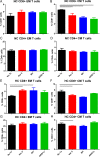
Fig 4. Peptide stimulation of splenocytes from apoE-/- mice fed normal chow or high fat diet.
Splenocytes from male apoE-/- mice fed normal chow (NC, A-D) or high fat diet (HC, E-H) for 6 weeks were stimulated with 20μg/ml of individual peptides for 24 hours and assessed for CD44+CD62L(-) Effector Memory (EM) or CD44+CD62L+ Central Memory (CM) T cells. Gating strategy is as indicated in Fig 3. No Tx = no peptide treatment; Ker II = Keratin, type II; BH = Bleomycin Hydrolase. Bar over control and peptide treated column indicates P<0.05. Spleens from 2–3 mice were pooled per group in triplicates.
Fig 5
Fig 5. Cytokine expression of splenocytes from apoE-/- mice after peptide stimulation.
Splenocytes were stimulated for 24 hours with 20μg/ml of the selected peptides to assess cytokine response. Gating depicted (A) includes cell singlets and excludes non-viable cells. Size-gated cells were then selected for CD3+ T cells, sub-grouped into CD8+ or CD4+ cells and assessed for IFN-γ or IL-10. No Tx = no peptide treatment; Ker II = Keratin, type II; BH = Bleomycin Hydrolase. Results are presented as percentage of CD8+ (B and C) or CD4+ (D and E) T cells. Spleens from 2–3 mice were pooled per group, (Ker II N = 3; No Tx, BH, ARID1a N = 4 each). Bar over columns indicate P<0.05.
Fig 6
Fig 6. CD4+FoxP3+ T regulatory cells in splenocytes of apoE-/- mice fed normal chow or high fat diet.
Splenocytes from apoE-/-FoxP3-GFP mice fed normal chow (NC; A) or high fat diet (HC; B) for 6 weeks were stimulated with 20μg/ml of individual peptides for 24 hours and assessed for FoxP3+ CD4+ T cells based on GFP fluorescence. No Tx = no peptide treatment; Ker II = Keratin, type II; BH = Bleomycin Hydrolase. Spleens from 2–3 mice were pooled per group, N = 3 each. Bar over columns indicate P<0.01.
Fig 7
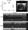
Fig 7. Myocardial infarction in apoE-/- mice.
A subgroup of mice fed normal chow were subjected to surgical myocardial infarction (MI) at 13 weeks of age and euthanized 6 weeks later. Control mice were not subjected to surgical manipulation, sham mice had surgery without coronary artery occlusion. Change in ejection fraction at 6 weeks post-surgery confirmed MI. Representative echocardiographic recording is shown Pre-MI and Post-MI.
Fig 8
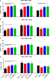
Fig 8. Peptide stimulation of splenocytes from apoE-/- mice subjected to MI.
Splenocytes from male apoE-/- mice 6 weeks after MI were stimulated with individual peptides for 24 hours. Cells from mice not subjected to surgery (Cont) or subjected to sham surgery were compared to mice subjected to MI. Cells were harvested and stained for flow cytometry for CD8+ (A and B) or CD4+ (C and D) EM and CM T cells. Gating strategy is as indicated in Fig 3. Bar over control and peptide treated column indicates statistical significance within respective group (Control P<0.01; Sham P<0.05; MI P<0.01). Spleens from 2–3 mice were pooled per group, N = 3 each. No Tx = no peptide treatment; Ker II = Keratin, type II; BH = Bleomycin Hydrolase.
Fig 9
Fig 9. Gating strategy used for flow cytometry of human PBMCs.
PBMCs were collected after 72 hours in culture with and without stimulation with Keratin 8 peptide. Positive control was stimulation with 0.5x T cell stimulation cocktail. Cells were collected and stained for flow cytometry. Cell singlets were selected and non-viable cells were excluded, followed by size-gating of viable cells. Cells were plotted on CD62L and CCR7 based on CD3+CD4+ cells or CD3+CD8+ cells. CD62L(-)CCR7(-) Effector cells were selected as T Effector Memory (TEM) or T Effector Memory RA+ (TEMRA) based on CD45RO/CD45RA stain.
Fig 10
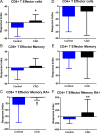
Fig 10. T Effector cell response to Keratin 8 peptide in CAD patient PBMC.
CD8+ (A-C) and CD4+ (D-F) Effector T cells in patient PBMC compared to control PBMC. T Effector Memory (B and E) or T Effector Memory RA+ (C and F) cells were based on CD45RO/CD45RA stain as depicted in the gating strategy as described in Fig 9. *P<0.05; †P = 0.07; **P<0.01. Control N = 15; CAD N = 17.
Fig 11
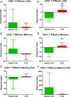
Fig 11. Effector T cell response in Stable CAD patients compared to ACS.
CD8+ (A-C) and CD4+ (D-F) Effector T cells in Stable CAD patient PBMC compared to ACS patient PBMC. Gating strategy is as depicted in Fig 9. **P<0.01. Stable CAD N = 7; ACS N = 10.
Fig 12
Fig 12. PBMC PD-1 mRNA expression in response to Keratin 8 stimulation.
PD-1 mRNA expression (A) in Control PBMC (N = 5) compared to CAD patient PBMC (N = 9) *P<0.05. CAD patients sub grouped (B) as Stable CAD (N = 4) or ACS (N = 5).

References

    1. Libby P, Tabas I, Fredman G, Fisher EA. Inflammation and its resolution as determinants of acute coronary syndromes. Circ Res. 2014;114(12): 1867–1879. 10.1161/CIRCRESAHA.114.302699 - DOI - PMC - PubMed
    1. Ridker PM, Everett BM, Thuren T, MacFadyen JG, Chang WH, Ballantyne C, et al. Antiinflammatory Therapy with Canakinumab for Atherosclerotic Disease. N Engl J Med. 2017. - PubMed
    1. Lu Y, Zhou S, Dreyer RP, Spatz ES, Geda M, Lorenze NP, et al. Sex Differences in Inflammatory Markers and Health Status Among Young Adults With Acute Myocardial Infarction: Results From the VIRGO (Variation in Recovery: Role of Gender on Outcomes of Young Acute Myocardial Infarction Patients) Study. Circ Cardiovasc Qual Outcomes. 2017;10(2): e003470 10.1161/CIRCOUTCOMES.116.003470 - DOI - PMC - PubMed
    1. Ridker PM. How Common Is Residual Inflammatory Risk? Circ Res. 2017;120(4): 617–619. 10.1161/CIRCRESAHA.116.310527 - DOI - PubMed
    1. Van Vre EA, Bult H, Hoymans VY, Van T, V, Vrints CJ, Bosmans JM. Human C-reactive protein activates monocyte-derived dendritic cells and induces dendritic cell-mediated T-cell activation. Arterioscler Thromb Vasc Biol. 2008;28(3): 511–518. 10.1161/ATVBAHA.107.157016 - DOI - PubMed

Publication types

MeSH terms