. 2019 Aug;61(2):219-231.
doi: 10.1165/rcmb.2018-0352OC.

Phosphorylation of GMFγ by c-Abl Coordinates Lamellipodial and Focal Adhesion Dynamics to Regulate Airway Smooth Muscle Cell Migration

Affiliations

Phosphorylation of GMFγ by c-Abl Coordinates Lamellipodial and Focal Adhesion Dynamics to Regulate Airway Smooth Muscle Cell Migration

Brennan D Gerlach et al. Am J Respir Cell Mol Biol. 2019 Aug.

Abstract

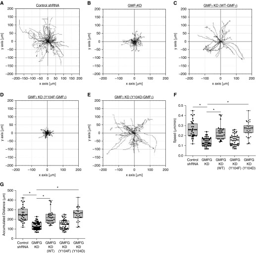
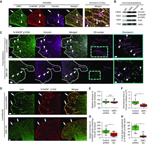
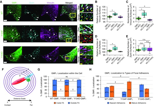
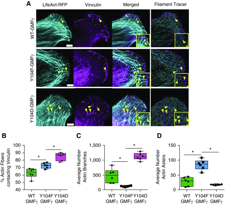
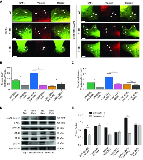
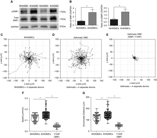
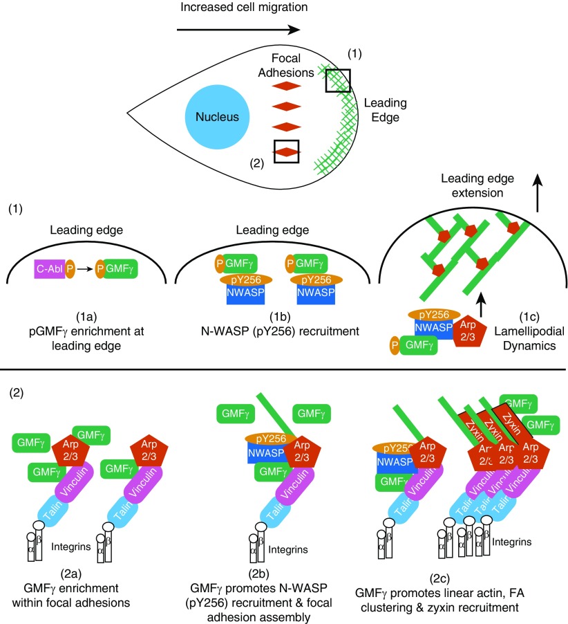
Airway smooth muscle cells require coordinated protrusion and focal adhesion dynamics to migrate properly. However, the signaling cascades that connect these two processes remain incompletely understood. Glia maturation factor (GMF)-γ has been implicated in inducing actin debranching and inhibiting nucleation. In this study, we discovered that GMFγ phosphorylation at Y104 regulates human airway smooth muscle cell migration. Using high-resolution microscopy coupled with three-dimensional object-based quantitative image analysis software, Imaris 9.2.0, phosphomimetic mutant, Y104D-GMFγ, was enriched at nascent adhesions along the leading edge where it recruited activated neural Wiskott-Aldrich syndrome protein (N-WASP; pY256) to promote actin-branch formation, which enhanced lamellipodial dynamics and limited the growth of focal adhesions. Unexpectedly, we found that nonphosphorylated mutant, Y104F-GMFγ, was enriched in growing adhesions where it promoted a linear branch organization and focal adhesion clustering, and recruited zyxin to increase maturation, thus inhibiting lamellipodial dynamics and cell migration. The localization of GMFγ between the leading edge and focal adhesions was dependent upon myosin activity. Furthermore, c-Abl tyrosine kinase regulated the GMFγ phosphorylation-dependent processes. Together, these results unveil the importance of GMFγ phosphorylation in coordinating lamellipodial and focal adhesion dynamics to regulate cell migration.

Keywords: actin cytoskeleton; airway smooth muscle; cell migration; protein phosphorylation.


Figures

Figure 1.
Figure 1.
(AE) GMFγ phosphorylation at Y104 regulates smooth muscle cell migration. Time-lapse microscopy was used to track human airway smooth muscle cells (HASMCs) expressing control shRNA and GMFγ shRNA, as well as cells transfected with wild-type (WT)-GMFγ, Y104F-GMFγ, and Y104D-GMFγ plasmids. Images were taken every 10 minutes for 16 hours. Migration plots generated by Image J plugin display migration patterns for each cell type. (F and G) Graphical comparisons represent the calculated speed and accumulated distance for each cell type. Two-tailed, one-way ANOVA with Tukey’s post hoc test was used (*P < 0.05; control shRNA n = 51, GMFγ knockdown (KD) n = 41, WT-GMFγ n = 27, Y104F-GMFγ n = 45, Y104D-GMFγ n = 31 n = pooled cell numbers from four human donors without asthma). GMFG = glia maturation factor γ.
Figure 2.
Figure 2.
Knockdown of GMFγ disrupts N-WASP (pY256) spatial distribution and reduces focal adhesion area. (A) HASMCs were plated on collagen-I–coated coverslips and immunostained for total GMFγ, N-WASP (pY256), and vinculin. Z-slice images were taken on a Zeiss LSM880 confocal microscope with the Fast Airyscan module using 488-, 561-, and 633-nm lasers. Scale bars: 5 μm and zoomed-in = 2 μm. Arrowheads point to focal adhesions and arrows point to the leading edge. Representative image from n = 10 individual cells. (B) Co-IP assay was performed on HASMCs using antibodies targeting endogenous GMFγ or vinculin. Co-IP samples were loaded and separated on an SDS-PAGE gel, where they were electrotransferred onto nitrocellulose paper and immunoblotted with primary antibodies for vinculin, N-WASP (pY256), Arp2, and GMFγ. Representative immunoblot from n = 4 independent experiments. (C) Control shRNA or GMFγ knockdown–expressing cells were immunostained for N-WASP (pY256) and vinculin. Z-slice images were taken on a Zeiss LSM880 confocal microscope with the Fast Airyscan module. Scale bar: 5 μm, zoomed-in = 2 μm. White line denotes the leading edge; arrowheads point to focal adhesions. (D) Control shRNA or GMFγ knockdown–expressing cells were immunostained for Arp2 and N-WASP (pY256). Arrows point to the leading edge. (EG) Imaris 9.1.2 software was used to 3D render spots (control shRNA = 18,433, GMFγ KD = 21,163) and surfaces (control shRNA = 1,993, GMFγ KD = 2,121) for quantitative analysis of vinculin number, area, and percent N-WASP spots contacting vinculin surfaces from 10 individual cells (Figure E3 and Methods). (H) Imaris 9.1.2 software was used to quantify the percent Arp2 spots (control shRNA = 12,660, GMFγ KD = 16,962) colocalized with N-WASP (pY256) spots (control shRNA = 10,879, GMFγ KD = 14,640) from 10 individual cells. Student’s t test was used (*P < 0.05). Arp2 = actin-related protein 2; Co-IP = co-immunoprecipitation; Ctrl = control; NHASMC = nonasthmatic HASMC; n.s. = not significant; N-WASP = neural Wiskott-Aldrich syndrome protein.
Figure 3.
Figure 3.
Phosphorylation at Y104 regulates focal adhesion clustering and GMFγ distribution. (A) GMFγ knockdown cells were transfected with WT, Y104F, or Y104D-GMFγ GFP-tagged plasmids, then fixed and immunostained for zyxin and vinculin. Arrowheads point to focal adhesions. Scale bars: white bars 50 μm and yellow bars 2 μm. (BE) Imaris 9.1.2 software was used to render GMFγ (WT = 49,304, Y104F = 45,524, Y104D = 27,178) spots, zyxin (WT = 2,228, Y104F = 3,390, Y104D = 755), and vinculin (WT = 4,482, Y104F = 7,751, Y104D = 3,800) surfaces from 10 individual cells to quantitate number and area of surfaces (Figure E3 and Methods). (F and G) Imaris 9.1.2 software was used to mask zyxin and vinculin surfaces followed by distance transformation algorithm to determine the percent of GMFγ inside or outside focal adhesions (FA). Distance transformation creates concentric circles around an object to measure distance based on the fluorescent intensity from other objects denoted by the distance scale (Methods). (H) Masked zyxin and vinculin channels were used to separate GMFγ localization within focal adhesions, and nascent adhesions contain only vinculin, whereas mature adhesions contain both vinculin and zyxin. A one-way ANOVA was used for statistical analysis with a Tukey’s post hoc test for between-group comparisons, *P < 0.05, #P < 0.05, and P < 0.05.
Figure 4.
Figure 4.
Actin architecture is regulated by GMFγ phosphorylation at Y104. (A) GMFγ knockdown cells were transfected with WT, Y104F, or Y104D-GMFγ and LifeAct–red fluorescent protein (RFP) (pseudocolored cyan) plasmids, fixed, and immunostained for vinculin. Images were taken using a Zeiss LSM880 confocal with the Fast Airyscan module. Scale bars: 5 μm, merged inset = 2 μm from n = 10 individual cells per expression plasmid. Arrowheads point to actin fibers and focal adhesions. (BD) Imaris software with the Filament Tracer package was used to trace actin fibers and analyze actin fibers contacting vinculin, as well as branch and aster morphology (Methods). Rendered vinculin (WT = 1,518, Y104F = 1,137, Y104D = 780) surfaces are in magenta, actin fibers (WT = 40,070, Y104F = 29,600, Y104D = 31,064) are in cyan, and “actin asters” or beginning points are labeled yellow. A one-way ANOVA with a Tukey’s post hoc test for between-group comparisons was used for statistical analysis. *P < 0.05.
Figure 5.
Figure 5.
Myosin activation regulates the recruitment of GMFγ to focal adhesions. (A) GMFγ knockdown cells were transfected with WT, Y104F, or Y104D-GMFγ plasmids overnight. Cells were then trypsinized and replated onto collagen-I–coated coverslips for 2 hours. After 2 hours, cells were treated with 20 μM blebbistatin for 15 minutes, then fixed and immunostained for vinculin. Scale bar = 10 μm; arrowheads point to focal adhesions. (B and C) Imaris 9.1.2 software was used to render vinculin (WT = 1,119, Y104F = 1,801, Y104D = 1,827) surfaces and GMFγ spots (WT = 14,916, Y104F = 14,475, Y104D = 10,723) for quantification of vinculin area and percent GMFγ spots contacting vinculin surfaces. Student’s t test was used for statistical analysis comparing no treatment to blebbistatin for each individual mutant GMFγ (n = 10 cells). *P < 0.05. (D) HASMCs were grown to confluence in 60-mm cell culture–treated dishes, then subjected to 20 μM (−/−) blebbistatin treatment for 15 minutes. Cells were harvested using 1× SDS sample buffer containing 1× protease and phosphatase inhibitor, scraped, and boiled for 5 minutes. Samples were run on SDS-PAGE, then electrotransferred onto nitrocellulose paper and immunoblotted for c-Abl tyr-412, total c-Abl, GAPDH, pMLC, MLC, GMFγ tyr-104, and total GMFγ. Western blots from n = 4 individual experiments were imaged using the GE Amersham 600 and analyzed using IQTL software. (E) Quantification of phospho:total protein ratio normalized to GAPDH was graphed. Student’s t test was used to compare the effect of blebbistatin treatment. *P < 0.05. Bleb = blebbistatin; c-ABL = a nonreceptor tyrosine kinase.
Figure 6.
Figure 6.
Phosphorylated GMFγ is upregulated in asthmatic HASMCs (AHASMCs) and contributes to its enhanced migratory phenotype. (A) Normal HASMC and airway smooth muscle cells isolated from five different donors with asthma were grown to confluence in 60-mm cell culture–treated dishes, then harvested with 1× SDS sample buffer containing 1× protease and phosphatase inhibitor, scraped, and boiled for 5 minutes. Samples were run on SDS-PAGE, then electrotransferred onto nitrocellulose paper and immunoblotted for phospho-GMFγ (custom antibody; Methods), total GMFγ, and GAPDH. Western blots were imaged using the GE Amersham 600 and analyzed using IQTL software. (B) Quantification of phospho:total protein ratio normalized to GAPDH was graphed. Student’s t test was used to compare normal versus asthma GMFγ expression. *P < 0.05. (CE) Cells were serum starved overnight then replated onto collagen-I–coated six-well dishes in 10% FBS medium. Time-lapse microscopy was used to track NHASMC and AHASMC migration with a start time 2 hours after seeding. Images were taken every 10 minutes for 16 hours. Migration plots were generated using an ImageJ plugin to trace individual cells migratory pattern. (F and G) Graphical comparisons represent the calculated speed and accumulated distance for each cell type. Two-tailed, one-way ANOVA with Tukey’s post hoc test was used (*P < 0.05; NHASMC n = 45, AHASMC n = 57, Y104F-GMFγ–expressing AHASMC n = 49; n represents pooled cell numbers from four donors without and five human donors with asthma).
Figure 7.
Figure 7.
Model: phosphorylation state of GMFγ dictates its localization and functionality to regulate cell migration. (1a) At the leading edge, cellular cues trigger the enrichment of phosphorylated GMFγ. (1b) There, phospho-GMFγ recruits N-WASP (pY256) to the leading edge to enhance actin reorganization through Arp2/3 activation. (1c) Increased actin remodeling leads to increased protrusion extension and enhances lamellipodial dynamics. (2a) Upon myosin activation, nonphosphorylated GMFγ becomes enriched within focal adhesions, which includes talin and integrins, as well as many other proteins. (2b) Nonphosphorylated GMFγ recruits N-WASP (pY256) and increases linear actin formation and focal adhesion assembly. (2c) Nonphosphorylated GMFγ promotes actin reorganization, focal adhesion clustering, and recruitment of zyxin to enhance focal adhesion maturation. Sustained mechanical tension will increase c-Abl activation within focal adhesions, leading to phosphorylation of GMFγ, thus liberating it from Arp2/3 and returning GMFγ to the leading edge.

Comment in

References

    1. Tang DD, Gerlach BD. The roles and regulation of the actin cytoskeleton, intermediate filaments and microtubules in smooth muscle cell migration. Respir Res. 2017;18:54. - PMC - PubMed
    1. Salter B, Pray C, Radford K, Martin JG, Nair P. Regulation of human airway smooth muscle cell migration and relevance to asthma. Respir Res. 2017;18:156. - PMC - PubMed
    1. Gizycki MJ, Adelroth E, Rogers AV, O’Byrne PM, Jeffery PK. Myofibroblast involvement in the allergen-induced late response in mild atopic asthma. Am J Respir Cell Mol Biol. 1997;16:664–673. - PubMed
    1. Kaminska M, Foley S, Maghni K, Storness-Bliss C, Coxson H, Ghezzo H, et al. Airway remodeling in subjects with severe asthma with or without chronic persistent airflow obstruction. J Allergy Clin Immunol. 2009;124:45–51.e1–4. - PubMed
    1. Krause M, Gautreau A. Steering cell migration: lamellipodium dynamics and the regulation of directional persistence. Nat Rev Mol Cell Biol. 2014;15:577–590. - PubMed

Publication types

MeSH terms