. 2019 Sep:82:54-70.
doi: 10.1016/j.matbio.2019.02.004. Epub 2019 Feb 24.

p16Ink4a deletion in cells of the intervertebral disc affects their matrix homeostasis and senescence associated secretory phenotype without altering onset of senescence

Affiliations

p16Ink4a deletion in cells of the intervertebral disc affects their matrix homeostasis and senescence associated secretory phenotype without altering onset of senescence

Emanuel J Novais et al. Matrix Biol. 2019 Sep.

Abstract

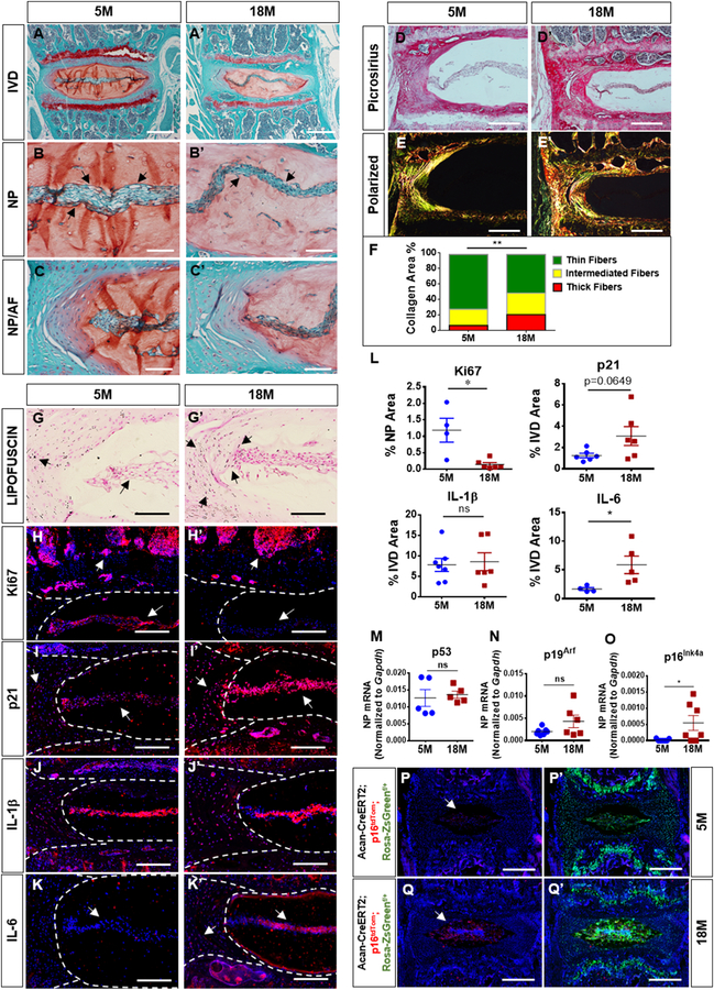
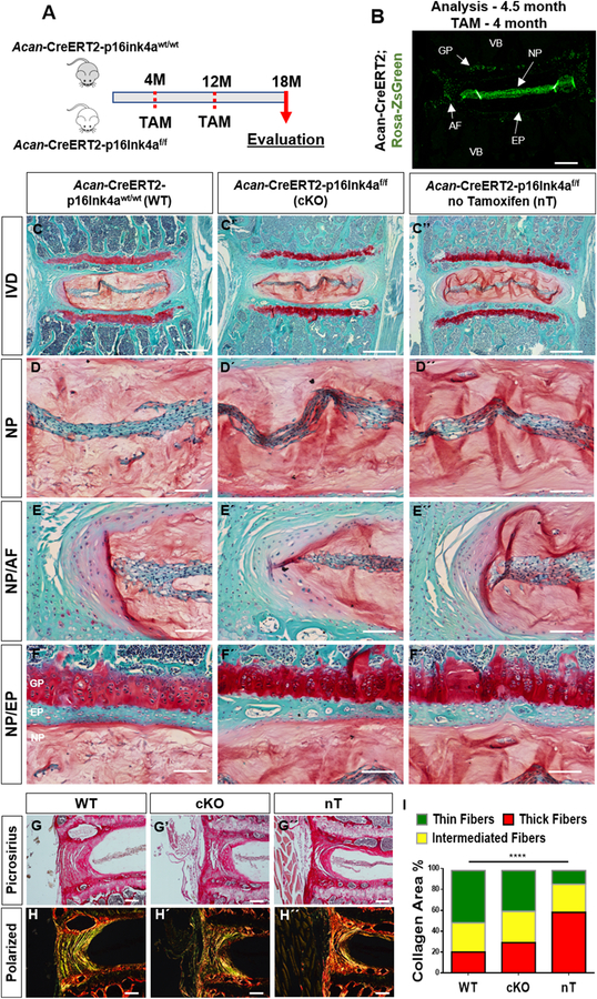
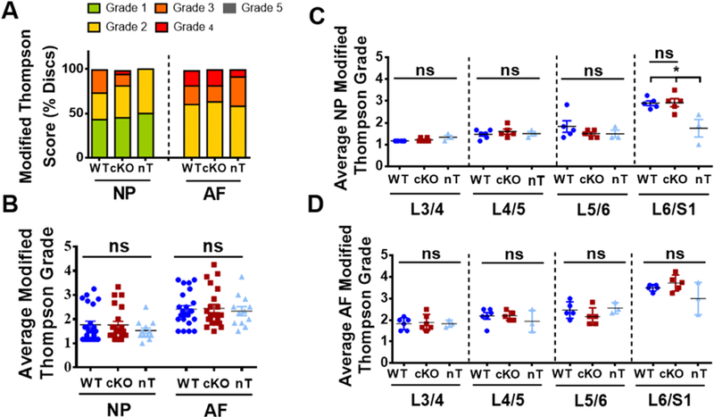
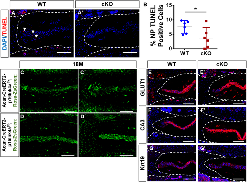
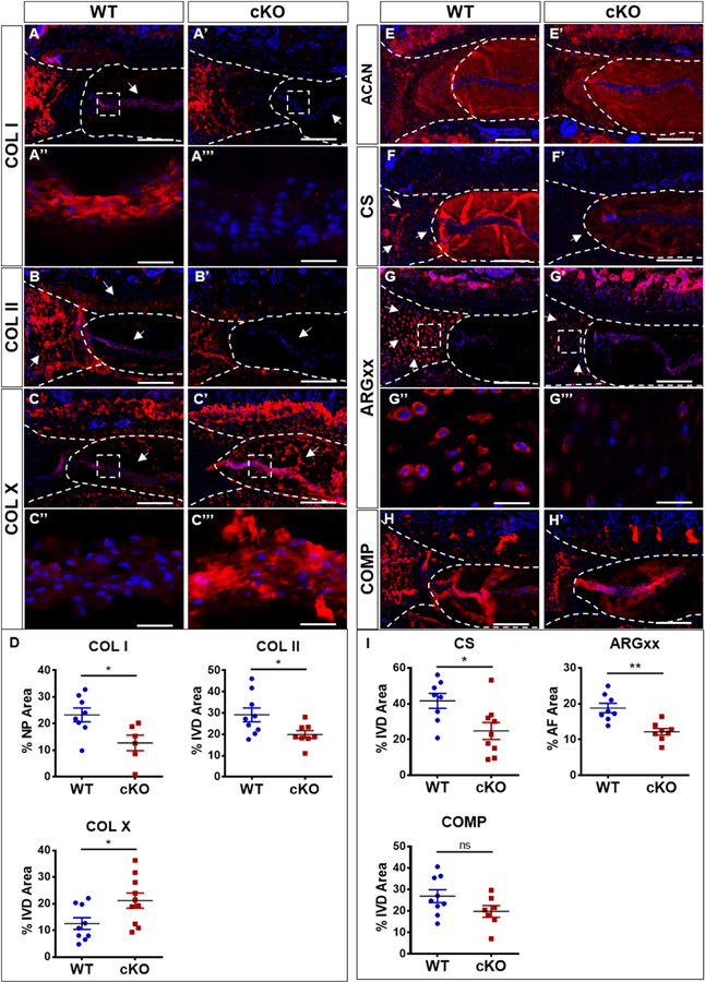
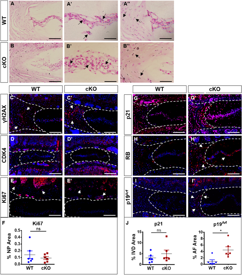
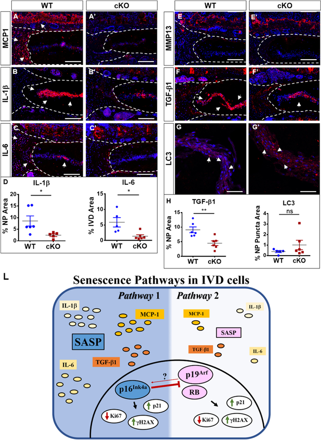
Intervertebral disc degeneration is an important contributor to chronic low back and neck pain. Although many environmental and genetic factors are known to contribute to disc degeneration, age is still the most significant risk factor. Recent studies have shown that senescence may play a role in age-related disc degeneration and matrix catabolism in humans and mouse models. Clearance of p16Ink4a-positive senescent cells reduces the degenerative phenotype in many age-associated diseases. Whether p16Ink4a plays a functional role in intervertebral disc degeneration and senescence is unknown. We first characterized the senescence status of discs in young and old mice. Quantitative histology, gene expression and a novel p16tdTom reporter mice showed an increase in p16Ink4a, p21 and IL-6, with a decrease in Ki67 with aging. Accordingly, we studied the spinal-phenotype of 18-month-old mice with conditional deletion of p16Ink4a in the disc driven by Acan-CreERT2 (cKO). The analyses of discs of cKO and age-matched control mice showed little change in cell morphology and tissue architecture. The cKO mice exhibited changes in functional attributes of aggrecan as well as in collagen composition of the intervertebral disc. While cKO discs exhibited a small decrease in TUNEL positive cells, lineage tracing experiments using ZsGreen reporter indicated that the overall changes in cell fate or numbers were minimal. The cKO mice maintained expression of NP-cell phenotypic markers CA3, Krt19 and GLUT-1. Moreover, in cKO discs, levels of p19Arf and RB were higher without alterations in Ki67, γH2AX, CDK4 and Lipofuscin deposition. Interestingly, the cKO discs showed lower levels of SASP markers, IL-1β, IL-6, MCP1 and TGF-β1. These results show that while, p16Ink4a is dispensable for induction and maintenance of senescence, conditional loss of p16Ink4a reduces apoptosis, limits the SASP phenotype and alters matrix homeostasis of disc cells.

Keywords: Aggrecan; Aging; Extracellular matrix; Ink4a; Intervertebral disc degeneration; Mouse models; Nucleus pulposus; SASP; Senescence; p16; p19.


Conflict of interest statement

Conflicts of Interest:

Nothing to disclosure.

Figures

Figure 1
Figure 1. Senescence and p16Ink4a expression increase with age. (A-C’)
18-month-old mice showed a decrease in vacuoles and cell band width (arrows) and comparable features of the NP/AF junction. (D-F) Picrosirius red staining (D-D’) and quantitative polarized imaging (E, E’) showed a decrease in thin collagen fibers along with an increase of medium and thick fibers compare to 5-month-old mice (F). P < 0.05, χ2 test, N = 4–6 mice/group, 4 discs/animal. (G-G’) Sudan-Black-B staining showed an increase in Lipofuscin aggregates deposition in NP and AF of old mice (arrows). (H-I’) There was a significant decrease in Ki67 and an increase of p21 expression in all disc compartments (arrows). (J-J’) Analysis of SASP showed comparable levels of IL-1β with aging. (K-K’) IL-6 expression was significantly higher in the older mice. (L) Quantification of % positive staining for KI67, p21, IL-1β and IL-6 expression. (M-N) p53 and p19Arf mRNA levels were similar between 5-and 18-month old mice(O) p16Ink4amRNA levels were significantly higher in 18-month old mice compared to 5-month old animals.(P-Q’) Acantm1(cre/ERT2)Crm; p16tdTom;Rosa-ZsGreen 18-month-old mice showed robust expression of p16Ink4a (tdTOM/Red) in the NP with strong colocalization with Aggrecan-Cre positive cells (ZsGreen), p16Ink4a levels were low in 5-moth-old mice. For quantitative immunohistology and RNA analysis, Mann-Whitney test was used for comparing differences between the groups. NS = not significant; p ≤ 0.05 *; p ≤ 0.01 **; N=6 animals/genotype were analyzed. Scale bar A-A’, P-Q’ = 200 µm; Scale bar B-E’’, G-K’ = 50 µm.
Figure 2
Figure 2. Loss of p16Ink4a in the disc does not protect mice from age-dependent degeneration.
(A) Schematic showing protocol for generation of experimental and control mice. Analysis was performed using tamoxifen treated 18-month-old wild-type (Acan-CreERT2-p16Ink4a wt/wt) and p16Ink4a conditional knock-out (Acan-CreERT2-p16Ink4a f/f) and untreated (Acan-CreERT2-p16Ink4af/f) animals. (B) Specificity of Acan-CreERT2 for targeting different compartments within the intervertebral disc in skeletally mature mice is shown. Acan-Cre showed a high recombination in NP, inner AF, endplate (EP) as well as growth plate (GP), when tamoxifen was administered at 4-month. (C-F’’) There are no noticeable differences in overall intervertebral disc (IVD) architecture or cellular morphology in the tamoxifen treated wild-type (WT) (C-F), conditional knock-out (cKO) (C’-F’), and animals without tamoxifen-treatment (nT) (C’’-F’’) groups as shown by representative histological images. Higher magnification images of NP tissue (D-D”) and tissue interfaces between NP/AF (E-E’’) and NP/EP (F-F’’) show comparable cell morphology and tissue architecture between all the groups. Picrosirius red staining of discs showed similar collagen content among the groups (G-G’’). There were no differences in collagen organization (H-H’’) and distribution of collagen fibers thickness (I) between the WT and cKO mice as seen by quantitative polarized microscopy. Non-treated animals showed higher content of mature collagen than tamoxifen treated WT and cKO animals. P < 0.05, χ2 test, N = 6 mice/group, 4 discs/animal. Scale bar B-C” and G-H´´= 200 µm; Scale bar D-F” = 50 µm.
Figure 3
Figure 3. Loss of p16Ink4a in the disc does not protect mice from age-dependent degeneration.
(A) Modified Thompson Grading showed no differences in the distribution of histopathological grades for both NP and AF amongst the groups. (B) The average NP and AF grades were similar for WT, cKO and nT animals. (C, D) Level-by-level analysis showed similar average grades for both NP (C) and AF (D) across the four lumbar levels L¾, L4/5, L5/6 and L6-S1 amongst all groups except for the average NP grade at L6/S1, where untreated animals showed a lower grade of degeneration than tamoxifen treated groups. 4 lumbar discs/mouse, 6 mice for WT and cKO and 3 mice for nT groups were analyzed for comparisons. Kruskal-Wallis test followed by Dunn’s multiple comparison test was used to test differences between groups showing average Thompson grading data. χ2 test was used to analyze differences between groups showing distribution of percent-degenerated-discs. NS = not significant; p ≤ 0.05 *.
Figure 4
Figure 4. p16Ink4a cKO mice have decreased NP cell death without significant altering cell number and phenotype.
(A-A’’) TUNEL assay showed a decrease in number of dying cells (arrows) in NP compartment of the cKO mouse. (B) There was a decrease in TUNEL positive cells in cKO mice. t-test for normally distributed data, Mann-Whitney test for non-normally distributed data, Shapiro-Wilk normality test was done to check the distribution. NS = not significant; p ≤ 0.05 *; p ≤ 0.01 **; N=6 animals/genotype were analyzed. (C, D’) Fate mapping of disc cells in p16Ink4a cKO and control mice. Tamoxifen treatment of mice (at 4 and 12-months) induced Cre-recombinase activity in aggrecan expressing cells resulting in simultaneous marking of cells by ZsGreen with or without deletion of p16Ink4a. Analysis of these mice at 18-months showed that almost all cells in NP, inner AF and CEP were ZsGreen positive. Cell numbers of ZsGreen positive cells in tissue compartments were comparable between cKO and control animals. (E-G’) Characterization of NP phenotypic markers in p16Ink4a cKO mice. Localization and expression levels of glucose transporter 1 (GLUT1), carbonic anhydrase 3 (CA3) and Keratin19 (Krt19) in the NP were comparable between control and cKO animals. Mann-Whitney test was used for comparing differences between the groups. NS = not significant; p ≤ 0.05 *; p ≤ 0.01 **; N=6 animals/genotype were analyzed. Scale bar = 200µm.
Figure 5
Figure 5. p16Ink4a cKO mice alter aggrecan functionality and expression of major extracellular matrix collagens of the disc.
(A-B’) cKO mice show decreased levels of collagen I and collagen II expression in the NP. (C-D) Collagen X was expressed in both groups with an increasing expression in cKO. (E-E) Analysis of aggrecan (ACAN) and its functional attributes. Expression of ACAN core protein was similar between the two genotypes (E-E’). Levels of chondroitin sulfate (CS) in NP (F-F’, I) and aggrecanases generated degradation product, ARGxx in AF were lower cKO group (G-G’’’, I). (H-I’). Mann-Whitney test was used for comparing differences between the groups. NS = not significant; p ≤ 0.05 *; p ≤ 0.01 **. N = 6 animals/genotype, 1–2 discs per animal, were analyzed. Scale bar = 200 µm (A-A’, B-C’, E-G’, H-H’ ) and 20 µm (A’’-A’’’, C’’-C’’’, G’’-G’’).
Figure 6
Figure 6. p16Ink4a cKO shows activation of p19Arf and RB without affecting the prevalence of senescent cells in the intervertebral disc.
(A-B’’) Sudan-Black-B staining showed comparable lipofuscin aggregates in WT and cKO mice. (C-F) There were no discernable changes in γH2AX or CDK4 and KI67 expression levels in the discs of the cKO and WT mice. (G-G’, J) p21 was expressed in all disc compartments in cKO and control animals. (H-H’) RB was higher expressed in overall disc of cKO mice. (I-J) cKO mice showed pronounced increase in p19Arf staining in the cells of inner AF and GP cells. For quantitative analysis, Mann-Whitney test was used for comparing differences between the groups. NS = not significant; p ≤ 0.05 *; N=6 animals/genotype were analyzed. Scale bar = 200 µm.
Figure 7
Figure 7. p16Ink4a cKO exhibit decreased expression of major regulators of senescence-associated secretory phenotype (SASP).
(A-C’) cKO animals showed a pronounced decrease in MCP1 (A, A’), IL1β (B, B’) and IL-6 (C, C’) staining compared to control animals (D). (E-E’) There were comparable levels of MMP13 expression between cKO mice and the WT. (F-F’, H) TGF-β1 expression was decreased in cKO discs. Autophagy analysis showed no differences between LC3 Puncta between WT and cKO (G-H). Mann-Whitney test was used for comparing differences between the groups. NS = not significant; p ≤ 0.05 *; N=6 animals/genotype were analyzed. Scale bar = 200 µm. (L) Schematic summarizing contribution of p16Ink4a, p19Arf and RB for senescence status and SASP maintenance in the intervertebral cells.

References

    1. Huang Y-C, Urban JPG, Luk KDK, Intervertebral disc regeneration: do nutrients lead the way?, Nat. Rev. Rheumatol 10 (2014) 1–6. doi: 10.1038/nrrheum.2014.91. - DOI - PubMed
    1. Choi H, Merceron C, Mangiavini L, Seifert EL, Schipani E, Shapiro IM, V Risbud M, Hypoxia promotes noncanonical autophagy in nucleus pulposus cells independent of MTOR and HIF1A signaling, Autophagy 12 (2016) 1631–1646. doi: 10.1080/15548627.2016.1192753. - DOI - PMC - PubMed
    1. Schoepflin ZR, Silagi ES, Shapiro IM, Risbud MV, PHD3 is a transcriptional coactivator of HIF-1α in nucleus pulposus cells independent of the PKM2-JMJD5 axis, FASEB J 31 (2017) 3831–3847. doi: 10.1096/fj.201601291R. - DOI - PMC - PubMed
    1. Choi H, Tessier S, Silagi ES, Kyada R, Yousefi F, Pleshko N, Shapiro IM, Risbud MV, A novel mouse model of intervertebral disc degeneration shows altered cell fate and matrix homeostasis, Matrix Biol 70 (2018) 102–122. doi: 10.1016/j.matbio.2018.03.019. - DOI - PMC - PubMed
    1. V Vo N, Hartman RA, Patil PR, V Risbud M, Kletsas D, Iatridis JC, Hoyland JA, Le Maitre CL, Sowa GA, James D, Molecular Mechanisms of Biological Aging in Intervertebral Discs, J Orthop Res 34 (2016) 1289–1306. doi: 10.1002/jor.23195. - DOI - PMC - PubMed

Publication types