. 2019 Feb 26;26(9):2509-2520.e4.
doi: 10.1016/j.celrep.2019.01.108.

Controlling Epithelial Polarity: A Human Enteroid Model for Host-Pathogen Interactions

Affiliations

Controlling Epithelial Polarity: A Human Enteroid Model for Host-Pathogen Interactions

Julia Y Co et al. Cell Rep. .

Abstract

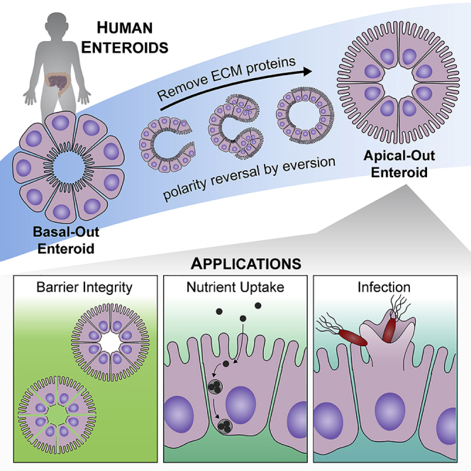
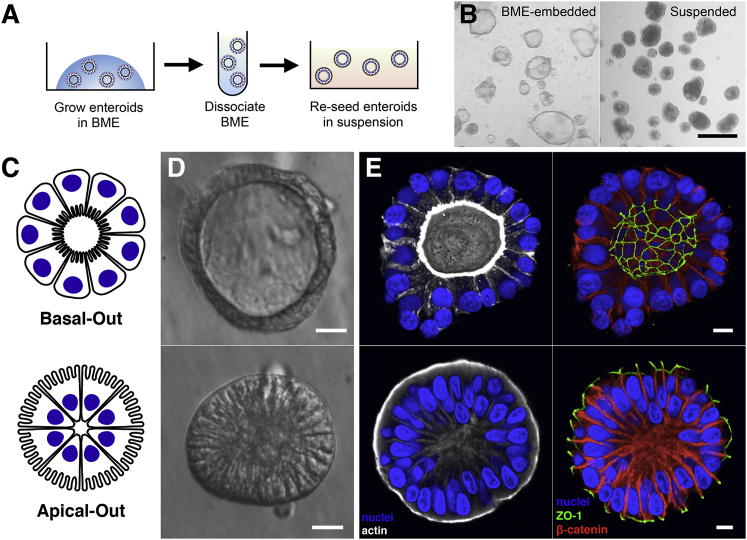
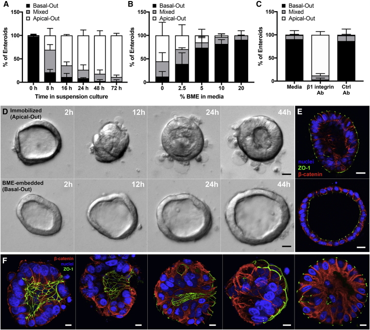
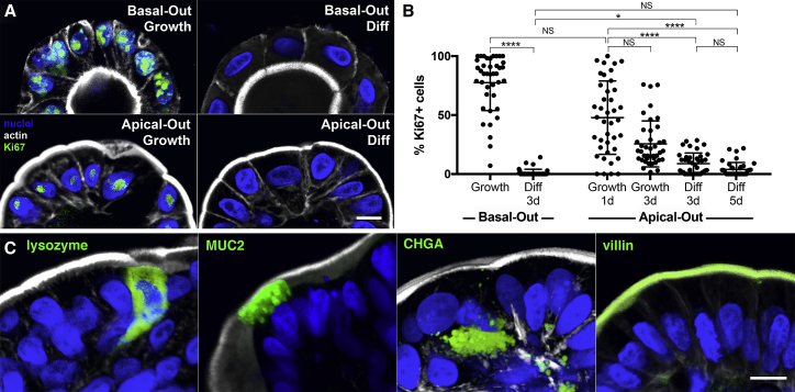
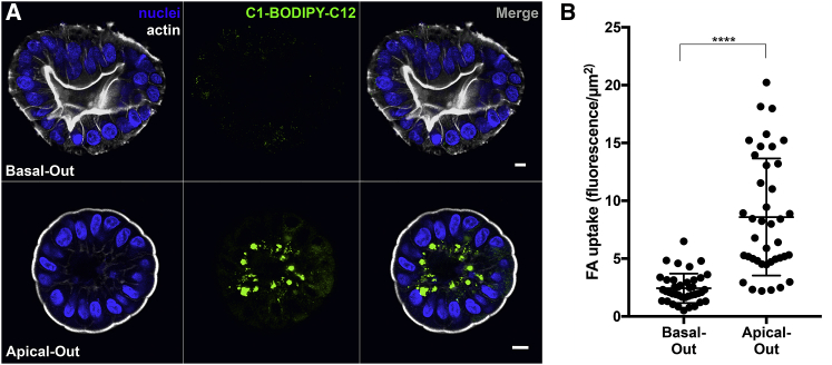
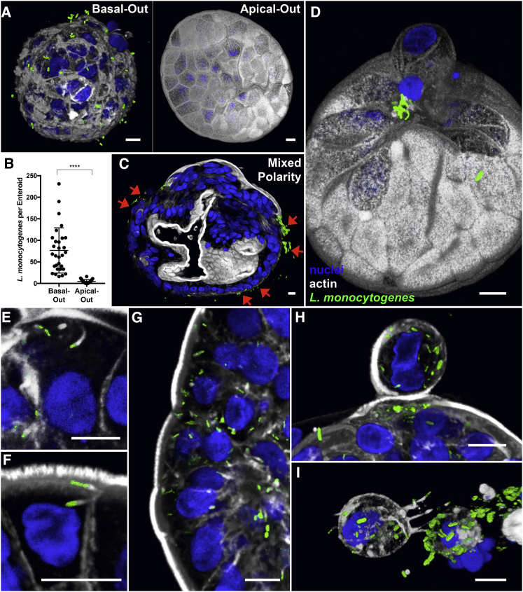
Human enteroids-epithelial spheroids derived from primary gastrointestinal tissue-are a promising model to study pathogen-epithelial interactions. However, accessing the apical enteroid surface is challenging because it is enclosed within the spheroid. We developed a technique to reverse enteroid polarity such that the apical surface everts to face the media. Apical-out enteroids maintain proper polarity and barrier function, differentiate into the major intestinal epithelial cell (IEC) types, and exhibit polarized absorption of nutrients. We used this model to study host-pathogen interactions and identified distinct polarity-specific patterns of infection by invasive enteropathogens. Salmonella enterica serovar Typhimurium targets IEC apical surfaces for invasion via cytoskeletal rearrangements, and Listeria monocytogenes, which binds to basolateral receptors, invade apical surfaces at sites of cell extrusion. Despite different modes of entry, both pathogens exit the epithelium within apically extruding enteroid cells. This model will enable further examination of IECs in health and disease.

Keywords: Listeria; Salmonella; apicobasal polarity; bacterial infection; epithelial organoids; gastrointestinal model; host-pathogen interactions; human enteroids; intestinal epithelial cells.


Figures

None
Graphical abstract
Figure 1
Figure 1
Enteroids in Suspension Culture Exhibit Apical-Out Polarity (A) Schematic for generation of suspended apical-out enteroids. (B) Images from a dissection microscope of BME-embedded enteroids (left) or suspended enteroids (right). Scale bar is 500 μm. (C–E) Basal-out enteroids and apical-out enteroids are (C) depicted schematically, (D) imaged using modulation contrast microscopy, and (E) imaged using confocal microscopy. Nuclei in blue, actin in white, ZO-1 in green, and β-catenin in red are shown. Scale bars are 10 μm. See also Figures S1, S2, and S4.
Figure 2
Figure 2
Characterization of Enteroid Polarity Reversal (A) Enteroids were analyzed using confocal microscopy and quantified for percentage of basal-out, apical-out, or mixed polarity enteroids; n = 3 experiments. (B) Quantification of basal-out, apical-out, or mixed polarity enteroids in suspension culture with soluble BME; n = 3 experiments. (C) BME-embedded enteroids were incubated in media alone or with β1-integrin function-blocking antibody or a control antibody for 1 day; n = 3 experiments. For (A)–(C), data represented are the means of each category with SD. (D) Time-lapse DIC microscopy of immobilized apical-out (top) or BME-embedded basal-out (bottom) enteroids as shown in Video S1. (E) Confocal microscopy of enteroids from time-lapse experiment. Nuclei in blue, ZO-1 in green, and β-catenin in red are shown. Scale bars are 20 μm. (F) Confocal microscopy of suspended enteroids at different stages of polarity reversal. Nuclei in blue, ZO-1 in green, and β-catenin in red are shown. Scale bars are 10 μm. See also Figures S3 and S4 and Video S1.
Figure 3
Figure 3
Dextran Diffusion Assay for Enteroid Epithelial Barrier Integrity (A) Apical-out enteroids exclude FITC-dextrans added to the media, demonstrating intact epithelial barrier integrity. (B) Treatment with 2 mM EDTA disrupts the epithelial barrier and results in diffusion of FITC-dextrans into the intercellular spaces and into the center of the spheroid. Enteroids imaged by DIC microscopy (left; scale bars are 50 μm), FITC-dextran fluorescence imaged by confocal microscopy (middle), and insets of center (right; scale bars are 10 μm) are shown. See also Figure S7.
Figure 4
Figure 4
Apical-Out Enteroids Proliferate and Differentiate (A) Basal-out enteroids and apical-out enteroids have proliferating cells when cultured in growth media (Growth), but not in differentiation media (Diff). (B) Quantification of proliferation marker Ki67 shows that both basal-out and apical-out enteroid cells proliferate less in differentiation media (Diff) than in growth media (Growth). The percentage of proliferating cells in apical-out enteroids in growth media decreases over time. Data represented are mean ± SD; n = 40 enteroids; p < 0.05; ∗∗∗∗p < 0.0001; NS, not significant using the Kruskal-Wallis test with Dunn’s multiple comparison test. (C) Markers for different epithelial cell types (lysozyme for Paneth cells, MUC2 for goblet cells, CHGA for entero-endocrine cells, and villin for enterocytes) are expressed in apical-out enteroids. Nuclei in blue and actin in white are shown. All scale bars are 10 μm. See also Figures S5 and S6.
Figure 5
Figure 5
Apical Absorption of Fatty Acids in Enteroids (A) Apical-out enteroids (bottom), but not basal-out enteroids (top), take up fluorescent fatty acid analog C1-BODIPY-C12 added to the extracellular media. Nuclei in blue and actin in white are shown. Scale bars are 10 μm. (B) Quantification of fatty acid analog (FA) uptake in apical-out and basal-out enteroids. Data represented are mean ± SD; n = 40 enteroids; p < 0.0001 using the Mann-Whitney U test.
Figure 6
Figure 6
S. Typhimurium Infection of Human Enteroids (A) S. Typhimurium-mCherry (red) at different stages of invasion of apical-out enteroids. (B) 3D confocal reconstructions of basal-out enteroids and apical-out enteroids infected with S. Typhimurium-mCherry for 1 hour. (C and D) Number of (C) invading bacteria and (D) actin ruffles per enteroid after 1 h of infection with S. Typhimurium-mCherry. Data represented are mean ± SD; n = 30 enteroids; p < 0.0001 using the Mann-Whitney U test. (E) S. Typhimurium-mCherry selectively invades the exposed apical surface (green arrows) of a mixed polarity enteroid. (F and G) 3D confocal reconstructions of S. Typhimurium-mCherry (F) within an epithelial cell in the process of extruding from the apical enteroid surface or (G) within a fully extruded cell after 6 h of infection. Nuclei in blue and actin in white are shown. All scale bars are 10 μm. See also Figure S7 and Video S2.
Figure 7
Figure 7
L. monocytogenes Infection of Human Enteroids (A) 3D confocal reconstruction of L. monocytogenes-GFP (green) attached to basal-out enteroids or apical-out enteroids after 15 min of infection. (B) Quantification of bacteria associated with basal-out or apical-out enteroids after 15 min of infection. Data represented are mean ± SD; n = 30 enteroids; p < 0.0001 using the Mann-Whitney U test. (C) L. monocytogenes-GFP selectively attaches to basal-out regions of a mixed polarity enteroid after 15 minutes of infection (red arrows). (D) 3D confocal reconstruction of a 15 minute infection shows that L. monocytogenes-GFP selectively attaches to apical-out enteroids at sites of cell extrusion. (E and F) After 6 h of infection, (E) intracellular L. monocytogenes-GFP are seen recruiting and polymerizing actin (white) into comet tails to move within the cytosol and (F) to invade neighboring cells. (G) Intracellular L. monocytogenes-GFP in patches of enteroid cells due to cell-to-cell spread. (H and I) In apical-out enteroids infected with L. monocytogenes-GFP for 6 h, bacteria exit the epithelium within (H) actively extruding cells or (I) completely extruded cells. Nuclei in blue and actin in white are shown. All scale bars are 10 μm.

References

    1. Bartfeld S., Clevers H. Organoids as model for infectious diseases: culture of human and murine stomach organoids and microinjection of Helicobacter pylori. J. Vis. Exp. 2015;105:e53359. - PMC - PubMed
    1. Bartfeld S., Bayram T., van de Wetering M., Huch M., Begthel H., Kujala P., Vries R., Peters P.J., Clevers H. In vitro expansion of human gastric epithelial stem cells and their responses to bacterial infection. Gastroenterology. 2015;148:126–136.e6. - PMC - PubMed
    1. Basak O., Beumer J., Wiebrands K., Seno H., van Oudenaarden A., Clevers H. Induced quiescence of Lgr5+ stem cells in intestinal organoids enables differentiation of hormone-producing enteroendocrine cells. Cell Stem Cell. 2017;20:177–190.e4. - PubMed
    1. Bishop A., House D., Perkins T., Baker S., Kingsley R.A., Dougan G. Interaction of Salmonella enterica serovar Typhi with cultured epithelial cells: roles of surface structures in adhesion and invasion. Microbiology. 2008;154:1914–1926. - PMC - PubMed
    1. Coburn B., Grassl G.A., Finlay B.B. Salmonella, the host and disease: a brief review. Immunol. Cell Biol. 2007;85:112–118. - PubMed

Publication types

MeSH terms