Review
. 2019 Feb 26;11(3):273.
doi: 10.3390/cancers11030273.

A Developmental Perspective on Paragangliar Tumorigenesis

Affiliations
Review

A Developmental Perspective on Paragangliar Tumorigenesis

Lavinia Vittoria Lotti et al. Cancers (Basel). .

Abstract

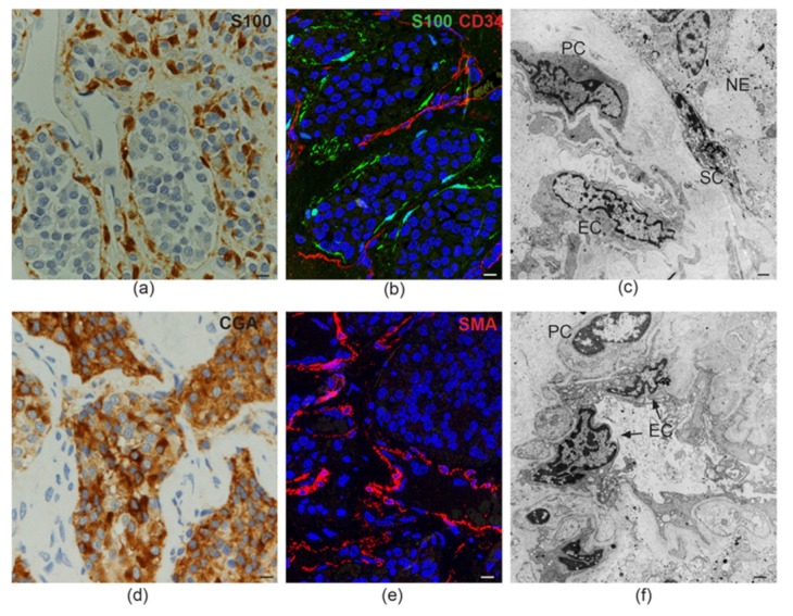
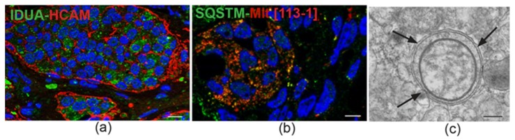
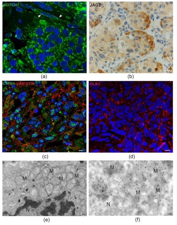
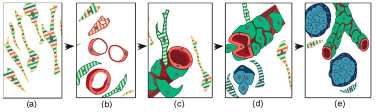
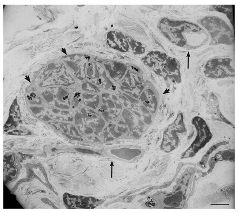
In this review, we propose that paraganglioma is a fundamentally organized, albeit aberrant, tissue composed of neoplastic vascular and neural cell types that share a common origin from a multipotent mesenchymal-like stem/progenitor cell. This view is consistent with the pseudohypoxic footprint implicated in the molecular pathogenesis of the disease, is in harmony with the neural crest origin of the paraganglia, and is strongly supported by the physiological model of carotid body hyperplasia. Our immunomorphological and molecular studies of head and neck paragangliomas demonstrate in all cases relationships between the vascular and the neural tumor compartments, that share mesenchymal and immature vasculo-neural markers, conserved in derived cell cultures. This immature, multipotent phenotype is supported by constitutive amplification of NOTCH signaling genes and by loss of the microRNA-200s and -34s, which control NOTCH1, ZEB1, and PDGFRA in head and neck paraganglioma cells. Importantly, the neuroepithelial component is distinguished by extreme mitochondrial alterations, associated with collapse of the ΔΨm. Finally, our xenograft models of head and neck paraganglioma demonstrate that mesenchymal-like cells first give rise to a vasculo-angiogenic network, and then self-organize into neuroepithelial-like clusters, a process inhibited by treatment with imatinib.

Keywords: angiogenesis; carotid body; mitochondria; neural crest; neurogenesis; paraganglioma; stem-like tumor cells; vasculogenesis; xenograft.


Conflict of interest statement

The authors declare no conflict of interest.

Figures

Figure 1
Figure 1
Tissue-like organization in paraganglioma. (a) Dark brown immunohistochemical staining for S100, a glial marker, highlights the sustentacular cell component surrounding the alveolar nests (“zellballen”) of neuroepithelial (“chief”) cells (avidin-biotin immunoperoxidase counterstained with hematoxylin and eosin, bar = 10 µm). (b) Immunofluorescence highlights thin sustentacular cells at the edges of neuroepithelial “zellballen,” identified by labeling with antibody to S100 (green). The endothelial lining of the capillaries in the surrounding stroma is labeled in red with CD34 (double immunofluorescence on semithin frozen section, bar = 10 µm). (c) Transmission electron micrograph showing the cytological features and topological relationships of the four main cell types that organize the paraganglioma microenvironment (endothelial cells, EC; pericytes, PC; sustentacular cells, SC; neuroepithelial cells, NE). Note the nuclear pleomorphism and similarities between chromatin patterns (bar = 2 µm). (d) Dark brown cytoplasmic staining for chromogranin A (CGA), a marker for neuroendocrine neoplasia, highlights neuroepithelial cell nests (“zellballen”) (avidin-biotin immunoperoxidase counterstained with hematoxylin and eosin, bar = 10 µm). (e) Immunofluorescence highlights the pericytic/mural cell component of the paraganglioma vasculature, identified by red labeling with antibody to smooth muscle actin (SMA, immunofluorescence on semithin frozen section, bar = 10 µm). (f) Ultrastructural cross section of a paraganglioma capillary showing the atypical cytological features of endothelial cells (EC) and pericytes (PC), two cell types whose roles in paragangliar tumorigenesis have been thus far scarcely considered (bar = 2 µm).
Figure 2
Figure 2
IDUA protein immunostaining and mitophagy in head and neck paraganglioma. (a) Immunofluorescence detects cytoplasmic IDUA protein labeling (green) in the neuroepithelial zellballen of paraganglioma. The zellballen are outlined in red by mainly peripheral labeling with antibody to HCAM/CD44, a surface stem cell marker that functions as a receptor for hyaluronan, a glycosaminoglycan degraded by the IDUA product (double immunofluorescence on semithin frozen section, bar = 10 µm). (b) Immunofluorescence highlights spots of colocalization (yellow) between anti-p62 antibody (Sequestosome-1, SQSTM, green) and anti-mitochondrial antibody 113-1 (ABCAM, red) (double immunofluorescence on semithin frozen section, bar = 10 µm). (c) Ultrastructural cross section of an autophagosome (indicated by arrows) containing a swollen mitochondrion, detected in the cytoplasm of a neuroepithelial paraganglioma cell (transmission electron micrograph, bar = 1 µm).
Figure 3
Figure 3
Notch pathway proteins in head and neck paraganglioma. (a) Semithin paraganglioma frozen section stained using immunofluorescence with an antibody that recognizes both membrane NOTCH1 and its active intracellular domain, NICD1 (green). In neuroepithelial cells, labeling is mainly concentrated in discrete cytoplasmic spots, suggesting mitochondrial localizations of NICD1. The adjacent endothelia (arrowheads) mainly reveal cell membrane NOTCH1 labeling (bar = 10 µm). (b) Immunohistochemical staining for the NOTCH ligand JAG2 is intense at the periphery of the neuroepithelial cell clusters, a typical location of the sustentacular cells (standard avidin-biotin immunoperoxidase counterstained with hematoxylin and eosin, bar = 10 µm). (c) Double immunofluorescence on semithin paraganglioma frozen section highlights punctate CTBP1 nuclear labeling in most cells. Red labeling identifies cells staining positive for vimentin, a mesenchymal marker (double immunofluorescence on semithin frozen section, bar = 10 µm). (d) Punctate membrane staining pattern (red) of the atypical NOTCH ligand DLK1 in paraganglioma cells (immunofluorescence on semithin frozen section, bar = 10 µm). (e) Electron micrograph of a neuroepithelial (“chief”) paraganglioma cell showing the accumulation of swollen mitochondria with disrupted cristae (M) next to the envelope of the nucleus (N) (bar = 1 µm). (f) Immunoelectron microscopic view of a similar ultrastructural field, showing dense NICD1 labeling of the perinuclear mitochondria with gold particles (ultrathin frozen section immunoelectronmicroscopy, bar = 1 µm).
Figure 4
Figure 4
NOTCH1 protein immunostaining in normal carotid body and in carotid body paraganglioma. (a) Low-power view of the fibroadipose tissue located at the carotid bifurcation, which contains paragangliar tissue immunostained (brown) with NOTCH1 antibody (avidin-biotin immunoperoxidase counterstained with hematoxylin and eosin, CA: carotid artery; FA: fibroadipose tissue; CB: carotid body; bar = 100 µm). (b) High-power view of the carotid body tissue immunostained (brown) with NOTCH1 antibody. Both capillary endothelia and neuroepithelial cells within “zellballen” are immunostained (avidin-biotin immunoperoxidase counterstained with hematoxylin and eosin, C: capillaries; ZB: “zellballen”; bar = 20 µm). (c) Paraganglioma tissue immunostained (brown) with NOTCH1 antibody. Ectatic capillaries and “zellballen” are immunostained (avidin-biotin immunoperoxidase counterstained with hematoxylin and eosin, C: capillaries; ZB: “zellballen”; bar = 25 µm).
Figure 5
Figure 5
Schematic representation of the histogenesis of head and neck paraganglioma, based on patient- and cell-derived xenograft models. The thin elongated stem-like cells (a), stabilized and expanded in paraganglioma cell cultures, co-express (stripes) multipotent stem/progenitor cell markers. In vivo, (b) these cells grow on autonomously synthesized extracellular matrix, develop intracytoplasmic vacuoles of increasing size (cells with red stripes only), and coalesce (uniformly red cells), giving rise to endothelial tubes (vasculogenesis via cytoplasmic hollowing). The endothelium then recruits adjacent stem-like cells (cells with green stripes only), which, after contact with the abluminal endothelial membranes (c), differentiate into mural cells (uniformly green). (d) The panel outlines two remarkable consequences of mural stabilization. First, mural impingement results in intraluminal endothelial intussusceptions, which divide the flow, giving rise to Y-shaped vascular ramifications (intussusceptive angiogenesis, a process detectable only with whole-mount confocal microscopy and/or transmission electron microscopy, as used in our study [56]). Secondly, vascular stabilization results in perivascular deposition of collagen IV [56], which supports the development of cell clusters with neural phenotype (cells with blue stripes only, then uniformly blue). As shown in (e), these clusters develop into “zellballen”-like neuroepithelial nests (uniform light blue), bound by spindle-shaped glia-like cells (uniform dark blue). Notably, while mesenchymal paraganglioma stem-like cells have normal mitochondria, paraganglioma tissue organization is associated with increasing mitochondrial dysfunction (swelling and loss of membrane potential), culminating in the neuroepithelial component. Original art by Giulio Pandolfelli, adapted and modified from Verginelli et al., 2018 [56].
Figure 6
Figure 6
Ultrastructural view of paraganglioma xenograft tissue. The electron micrograph, derived from a xenograft obtained by subcutaneous injection of an immortalized tympano-jugular paraganglioma cell line (PTJ64i), shows a tight neuroepithelial-like cell cluster (arrowheads, dark spots are lipofuscins) in the context of a vasculogenic tissue revealing endothelial-like cells with cytoplasmic hollowing and capillary-like structures (arrows) (bar = 5 µm).

Similar articles

Cited by

References

    1. Kho A.T., Zhao Q., Cai Z., Butte A.J., Kim J.Y., Pomeroy S.L., Rowitch D.H., Kohane I.S. Conserved mechanisms across development and tumorigenesis revealed by a mouse development perspective of human cancers. Genes Dev. 2004;18:629–640. doi: 10.1101/gad.1182504. - DOI - PMC - PubMed
    1. Krishna Priya S., Nagare R.P., Sneha V.S., Sidhanth C., Bindhya S., Manasa P., Ganesan T.S. Tumour angiogenesis-Origin of blood vessels. Int. J. Cancer. 2016;139:729–735. doi: 10.1002/ijc.30067. - DOI - PubMed
    1. Beachy P.A., Karhadkar S.S., Berman D.M. Tissue repair and stem cell renewal in carcinogenesis. Nature. 2004;432:324–331. doi: 10.1038/nature03100. - DOI - PubMed
    1. Levin M. Morphogenetic fields in embryogenesis, regeneration, and cancer: Non-local control of complex patterning. Biosystems. 2012;109:243–261. doi: 10.1016/j.biosystems.2012.04.005. - DOI - PMC - PubMed
    1. Simon M.C., Keith B. The role of oxygen availability in embryonic development and stem cell function. Nat. Rev. Mol. Cell Biol. 2008;9:285–296. doi: 10.1038/nrm2354. - DOI - PMC - PubMed

LinkOut - more resources