. 2019 Feb 26;11(3):277.
doi: 10.3390/cancers11030277.

Inhibition of NK Reactivity Against Solid Tumors by Platelet-Derived RANKL

Affiliations

Inhibition of NK Reactivity Against Solid Tumors by Platelet-Derived RANKL

Kim L Clar et al. Cancers (Basel). .

Abstract

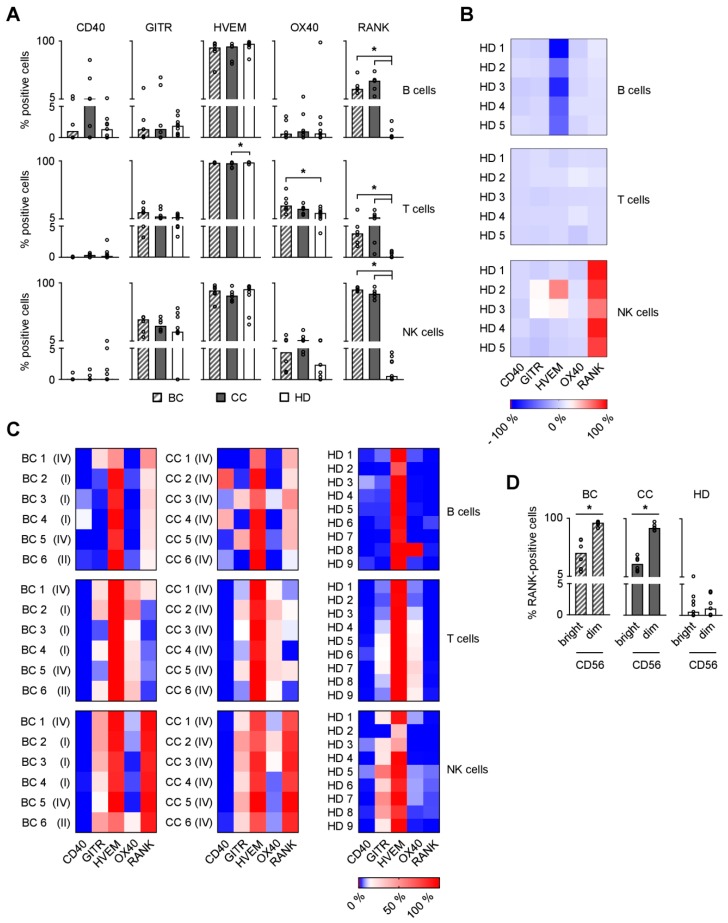
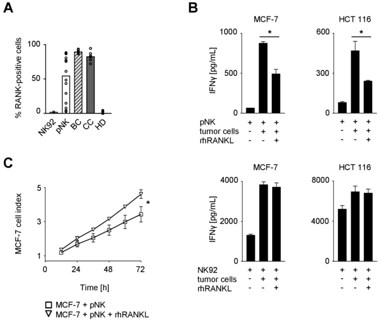
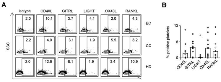
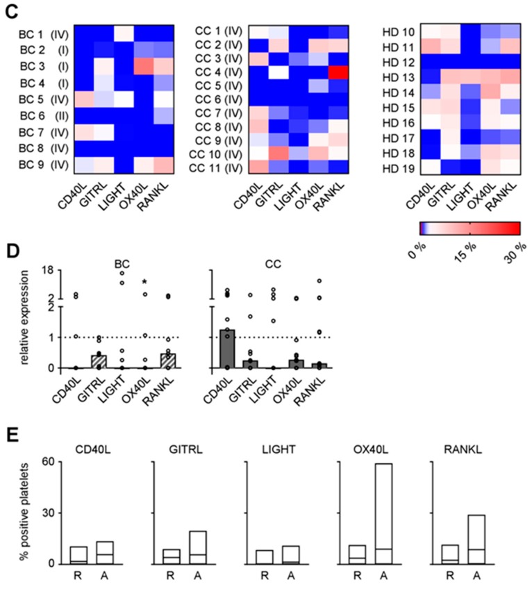
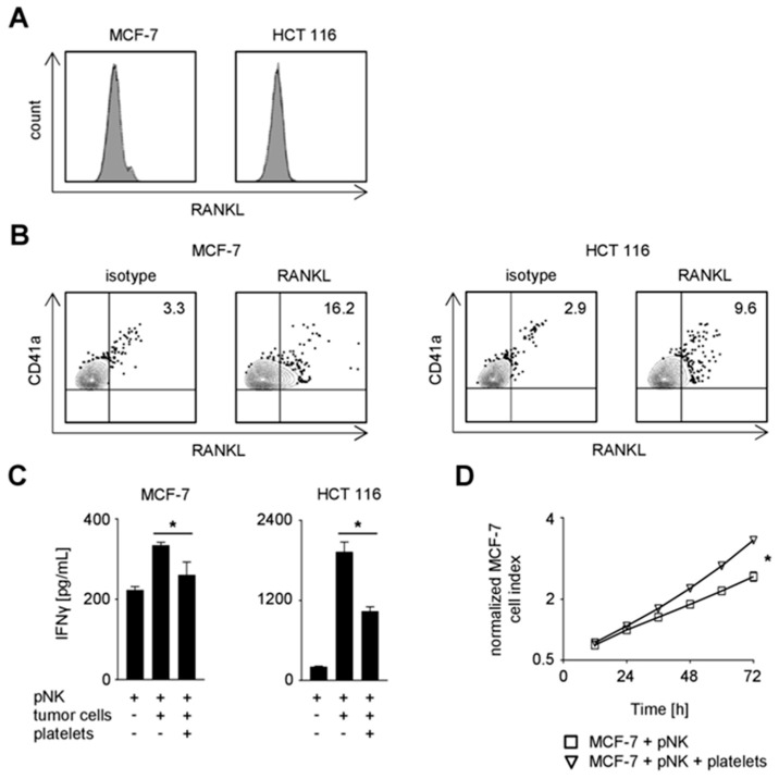
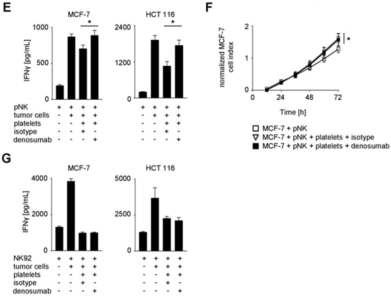
NK cells play an important role in tumor immunosurveillance. Their reactivity is governed by various activating and inhibitory surface receptors, which include several members of the TNF/TNF receptor family. For more than 50 years, it has been recognized that tumor immunosurveillance and in particular NK cell antitumor reactivity is largely influenced by platelets, but the underlying mechanisms remain to be fully elucidated. Here we report that upon activation, which reportedly occurs following interaction with cancer cells, platelets upregulate the TNF family member RANKL. Comparative analysis of the expression of RANK among different NK cell subsets and RANKL on platelets in cancer patients and healthy volunteers revealed a distinct malignant phenotype, and platelet-derived RANKL was found to inhibit the activity of normal NK cells against cancer cells. Notably, NK cell antitumor reactivity could be partially restored by application of denosumab, a RANKL-neutralizing antibody approved for treatment of benign and malignant osteolysis. Together, our data not only unravel a novel mechanism of tumor immune evasion mediated by platelets, but they also provide a functional explanation for the clinical observation that denosumab, beyond protecting from bone loss, may prolong disease-free survival in patients with solid tumors.

Keywords: NK cells; RANK/RANKL; cancer; denosumab; immune evasion; metastasis; platelets.


Conflict of interest statement

The authors declare no conflict of interest.

Figures

Figure 1
Figure 1
Expression of TNFR family molecules on PBMC subpopulations. (A,C,D) CD40, GITR, HVEM, OX40, and RANK surface expression on PBMC subpopulations from BC and CC patients and HD were investigated by flow cytometry (n = 6, 6, and 9, respectively). (A) The percentage of surface expression is indicated. (B) PBMC from five HD were freshly isolated or cultured without treatment for three days and CD40, GITR, HVEM, OX40, and RANK surface expression was determined by flow cytometry. Results were comparatively analyzed as follows: “percent surface expression of cultured PBMC” − “percent surface expression of freshly isolated PBMC”. The net modulation is depicted as heatmap. (C) Heatmap analysis of the surface expression profiles among PBMC of individual patients (disease stage as described in Table 1) and HD investigated. (D) The percentage of RANK surface expression on CD56bright and CD56dim NK cell subsets is displayed. (A,D) Median values of the respective group are depicted. Statistically significantly different results (p < 0.05) are indicated by *.
Figure 2
Figure 2
Expression of RANK and functional role of the RANK/RANKL axis in NK cell reactivity against solid tumors. (A) RANK surface expression on NK92 cells, pNK cells, and NK cells among PBMC from BC and CC patients and HD was investigated by flow cytometry (n = 1, 14, 6, 6, and 9, respectively). Median values of the respective group are depicted. (B) pNK (upper panel) or NK92 cells (lower panel) were cultured in the presence or absence of the indicated tumor cells and rhRANKL (125 ng/mL). IFNγ levels in culture supernatants were determined by ELISA after 24 h. (C) pNK cells were co-cultured with MCF-7 cells in the presence or absence of rhRANKL (125 ng/mL). The effect of NK cell reactivity on tumor cell proliferation/survival was assessed by xCELLigence RTCA for 72 h. Results are shown as electrical impedance signal (given as cell index). (B,C) Representative data of one experiment from a total of at least three with similar results are shown. Statistically significantly different results (p < 0.05) are indicated by *.
Figure 3
Figure 3
Expression of TNF family molecules on platelets. (A–D) CD40L, GITRL, LIGHT, OX40L, and RANKL surface expression on resting platelets from BC and CC patients and HD were investigated by flow cytometry after fixation with 2% paraformaldehyde (n = 9, 11, and 10, respectively). (A) Representative results obtained from BC and CC patients and HD are shown. (B) The percentage of surface expression on platelets from HD is indicated. (C) Heatmap analysis of the expression profiles among the platelets of individual patients (disease stage as described in Table 1) and HD investigated. (D) Relative surface expression on platelets from BC and CC patients compared to HD is depicted. For combined analysis, the median percentage of positive platelets obtained from HD was set to 1 for each individual TNF family molecule analyzed (dotted lines). (E) The percentage of CD40L, GITRL, LIGHT, OX40L, and RANKL surface expression on resting (R) or activated (exposure to thrombin for 1 min, A) platelets from HD was analyzed by flow cytometry after fixation with 2% paraformaldehyde (R, n = 10; A, n = 6). (B,D) Median values of the respective group are depicted. (D,E) Statistically significantly different results (p < 0.05) are indicated by *.
Figure 3
Figure 3
Expression of TNF family molecules on platelets. (A–D) CD40L, GITRL, LIGHT, OX40L, and RANKL surface expression on resting platelets from BC and CC patients and HD were investigated by flow cytometry after fixation with 2% paraformaldehyde (n = 9, 11, and 10, respectively). (A) Representative results obtained from BC and CC patients and HD are shown. (B) The percentage of surface expression on platelets from HD is indicated. (C) Heatmap analysis of the expression profiles among the platelets of individual patients (disease stage as described in Table 1) and HD investigated. (D) Relative surface expression on platelets from BC and CC patients compared to HD is depicted. For combined analysis, the median percentage of positive platelets obtained from HD was set to 1 for each individual TNF family molecule analyzed (dotted lines). (E) The percentage of CD40L, GITRL, LIGHT, OX40L, and RANKL surface expression on resting (R) or activated (exposure to thrombin for 1 min, A) platelets from HD was analyzed by flow cytometry after fixation with 2% paraformaldehyde (R, n = 10; A, n = 6). (B,D) Median values of the respective group are depicted. (D,E) Statistically significantly different results (p < 0.05) are indicated by *.
Figure 4
Figure 4
Functional role of platelet-derived RANKL in NK cell reactivity against solid tumors. (A) RANKL surface expression on the indicated tumor cells was investigated by flow cytometry. (BG) The indicated tumor cells were incubated in the presence or absence of platelets from HD. Coating was performed as described in the Materials and Methods section. (B) RANKL and CD41a surface expression on the indicated platelet-coated tumor cells was investigated by flow cytometry. (C) pNK cells were cultured in the presence or absence of the indicated tumor cells and platelets from HD. IFNγ levels in the culture supernatants were determined by ELISA after 24 h. (D) pNK cells were co-cultured with MCF-7 cells in the presence or absence of platelets from HD. The effect of NK cell reactivity on tumor cell proliferation/survival was assessed by xCELLigence RTCA for 72 h. Results are shown as electrical impedance signal (given as normalized cell index). Cell index was normalized after addition of NK cells to the tumor cells. (E) pNK cells were cultured in the presence or absence of the indicated tumor cells and platelets from HD. Where denoted, denosumab (10 µg/mL) or the respective isotype control was applied. IFNγ levels in the culture supernatants were determined by ELISA after 24 h. (F) pNK cells were treated as indicated in (E) and analyzed as in (D). (G) NK92 cells were treated and analyzed as described in (E). (CG) Representative data of one experiment from a total of at least three with similar results are shown. Statistically significantly different results (p < 0.05) are indicated by *.
Figure 4
Figure 4
Functional role of platelet-derived RANKL in NK cell reactivity against solid tumors. (A) RANKL surface expression on the indicated tumor cells was investigated by flow cytometry. (BG) The indicated tumor cells were incubated in the presence or absence of platelets from HD. Coating was performed as described in the Materials and Methods section. (B) RANKL and CD41a surface expression on the indicated platelet-coated tumor cells was investigated by flow cytometry. (C) pNK cells were cultured in the presence or absence of the indicated tumor cells and platelets from HD. IFNγ levels in the culture supernatants were determined by ELISA after 24 h. (D) pNK cells were co-cultured with MCF-7 cells in the presence or absence of platelets from HD. The effect of NK cell reactivity on tumor cell proliferation/survival was assessed by xCELLigence RTCA for 72 h. Results are shown as electrical impedance signal (given as normalized cell index). Cell index was normalized after addition of NK cells to the tumor cells. (E) pNK cells were cultured in the presence or absence of the indicated tumor cells and platelets from HD. Where denoted, denosumab (10 µg/mL) or the respective isotype control was applied. IFNγ levels in the culture supernatants were determined by ELISA after 24 h. (F) pNK cells were treated as indicated in (E) and analyzed as in (D). (G) NK92 cells were treated and analyzed as described in (E). (CG) Representative data of one experiment from a total of at least three with similar results are shown. Statistically significantly different results (p < 0.05) are indicated by *.

Similar articles

Cited by

References

    1. Gasic G.J., Gasic T.B., Stewart C.C. Antimetastatic effects associated with platelet reduction. Proc. Natl. Acad. Sci. USA. 1968;61:46–52. doi: 10.1073/pnas.61.1.46. - DOI - PMC - PubMed
    1. Gay L.J., Felding-Habermann B. Contribution of platelets to tumour metastasis. Nat. Rev. Cancer. 2011;11:123–134. doi: 10.1038/nrc3004. - DOI - PMC - PubMed
    1. Labelle M., Begum S., Hynes R.O. Direct signaling between platelets and cancer cells induces an epithelial-mesenchymal-like transition and promotes metastasis. Cancer Cell. 2011;20:576–590. doi: 10.1016/j.ccr.2011.09.009. - DOI - PMC - PubMed
    1. Placke T., Kopp H.G., Salih H.R. Modulation of Natural Killer Cell Anti-Tumor Reactivity by Platelets. J. Innate Immun. 2011;4:374–382. doi: 10.1159/000323936. - DOI - PubMed
    1. Nieswandt B., Hafner M., Echtenacher B., Mannel D.N. Lysis of tumor cells by natural killer cells in mice is impeded by platelets. Cancer Res. 1999;59:1295–1300. - PubMed

LinkOut - more resources