

Comparison of myocardial fibrosis quantification methods by cardiovascular magnetic resonance imaging for risk stratification of patients with suspected myocarditis
- PMID: 30813942
- PMCID: PMC6393997
- DOI: 10.1186/s12968-019-0520-0
Comparison of myocardial fibrosis quantification methods by cardiovascular magnetic resonance imaging for risk stratification of patients with suspected myocarditis
Abstract
Background: Although the presence of late gadolinium enhancement (LGE) using cardiovascular magnetic resonance imaging (CMR) is a significant discriminator of events in patients with suspected myocarditis, no data are available on the optimal LGE quantification method.
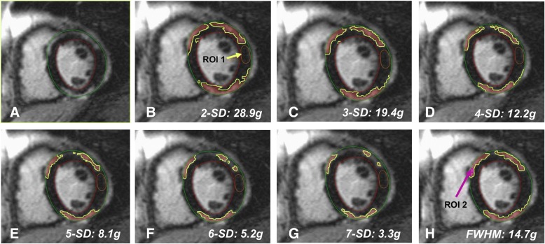
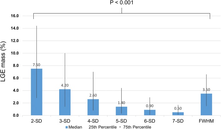
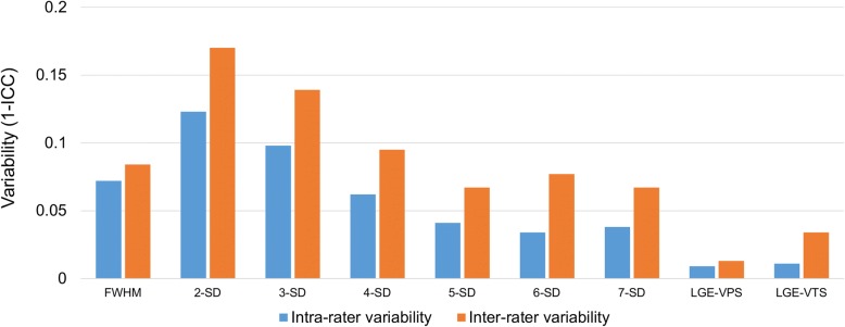
Methods: Six hundred seventy consecutive patients (48 ± 16 years, 59% male) with suspected myocarditis were enrolled between 2002 and 2015. We performed LGE quantitation using seven different signal intensity thresholding methods based either on 2, 3, 4, 5, 6, 7 standard deviations (SD) above remote myocardium or full width at half maximum (FWHM). In addition, a LGE visual presence score (LGE-VPS) (LGE present/absent in each segment) was assessed. For each of these methods, the strength of association of LGE results with major adverse cardiac events (MACE) was determined. Inter-and intra-rater variability using intraclass-correlation coefficient (ICC) was performed for all methods.
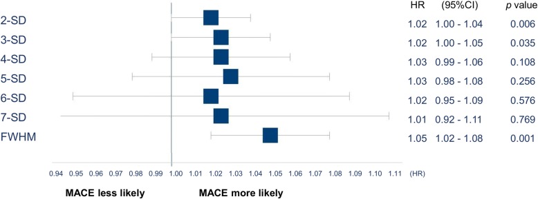
Results: Ninety-eight (15%) patients experienced a MACE at a medium follow-up of 4.7 years. LGE quantification by FWHM, 2- and 3-SD demonstrated univariable association with MACE (hazard ratio [HR] 1.05, 95% confidence interval [CI]:1.02-1.08, p = 0.001; HR 1.02, 95%CI:1.00-1.04; p = 0.001; HR 1.02, 95%CI: 1.00-1.05, p = 0.035, respectively), whereas 4-SD through 7-SD methods did not reach significant association. LGE-VPS also demonstrated association with MACE (HR 1.09, 95%CI: 1.04-1.15, p < 0.001). In the multivariable model, FWHM, 2-SD methods, and LGE-VPS each demonstrated significant association with MACE adjusted to age, sex, BMI and LVEF (adjusted HR of 1.04, 1.02, and 1.07; p = 0.009, p = 0.035; and p = 0.005, respectively). In these, FWHM and LGE-VPS had the highest degrees of inter and intra-rater reproducibility based on their high ICC values.
Conclusions: FWHM is the optimal semi-automated quantification method in risk-stratifying patients with suspected myocarditis, demonstrating the strongest association with MACE and the highest technical consistency. Visual LGE scoring is a reliable alternative method and is associated with a comparable association with MACE and reproducibility in these patients.
Trial registration number: NCT03470571 . Registered 13th March 2018. Retrospectively registered.
Keywords: CMR; Cardiovascular magnetic resonance imaging; FWHM; Full width half maximum; MACE; Myocarditis; Outcome; Quantification method; SD; Standard deviation.
Conflict of interest statement
Ethics approval and consent to participate
All study procedures were reviewed and approved by our Institutional Review Board at Brigham and Women’s Hospital, Harvard Medical School Boston in accordance with institutional guidelines. Given the retrospective nature of the current data spanning the past decade, obtaining informed consent from each patient was not logistically feasible, and a waiver for signing informed consent was obtained from by our Institutional Review Board. For patients who were followed-up by email or phone, signed informed consent is available.
Consent for publication
Not applicable
Competing interests
The authors declare that they have no competing interests.
Publisher’s Note
Springer Nature remains neutral with regard to jurisdictional claims in published maps and institutional affiliations.
Figures
References
-
- Cooper LT, Baughman KL, Feldman AM, et al. The role of endomyocardial biopsy in the management of cardiovascular disease: a scientific statement from the American Heart Association, the American College of Cardiology, and the European Society of Cardiology. Endorsed by the Heart Failure Society of America and the heart failure Association of the European Society of cardiology. J Am Coll Cardiol. 2007;50(19):1914–1931. doi: 10.1016/j.jacc.2007.09.008. - DOI - PubMed
Publication types
MeSH terms
Substances
Associated data
Grants and funding
LinkOut - more resources
Full Text Sources
Medical
