. 2019 Apr 19;294(16):6635-6644.
doi: 10.1074/jbc.RA119.007831. Epub 2019 Feb 27.

Structure-function analyses reveal that a glucuronoyl esterase from Teredinibacter turnerae interacts with carbohydrates and aromatic compounds

Affiliations

Structure-function analyses reveal that a glucuronoyl esterase from Teredinibacter turnerae interacts with carbohydrates and aromatic compounds

Jenny Arnling Bååth et al. J Biol Chem. .

Abstract

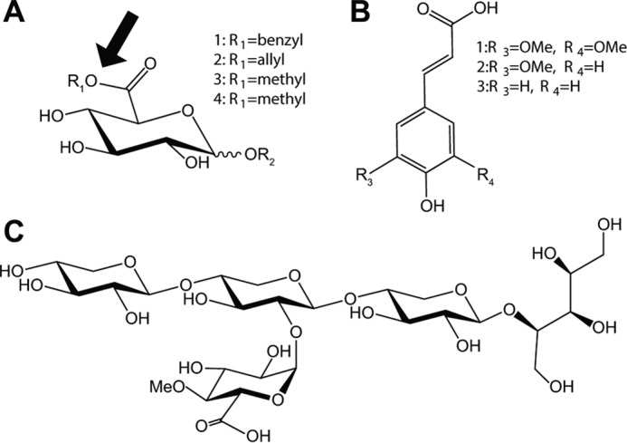
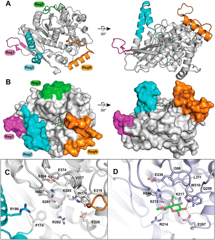
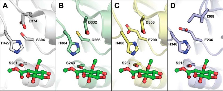
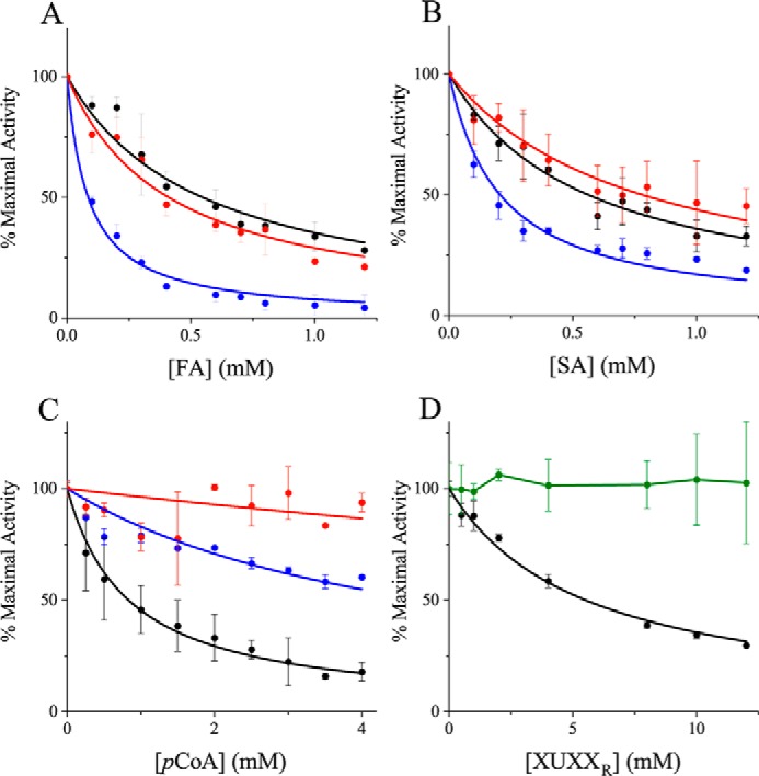
Glucuronoyl esterases (GEs) catalyze the cleavage of ester linkages found between lignin and glucuronic acid moieties on glucuronoxylan in plant biomass. As such, GEs represent promising biochemical tools in industrial processing of these recalcitrant resources. However, details on how GEs interact with their natural substrates are sparse, calling for thorough structure-function studies. Presented here is the structure and biochemical characterization of a GE, TtCE15A, from the bacterium Teredinibacter turnerae, a symbiont of wood-boring shipworms. To gain deeper insight into enzyme-substrate interactions, inhibition studies were performed with both the WT TtCE15A and variants in which we, by using site-directed mutagenesis, substituted residues suggested to have key roles in binding to or interacting with the aromatic and carbohydrate structures of its uronic acid ester substrates. Our results support the hypothesis that two aromatic residues (Phe-174 and Trp-376), conserved in bacterial GEs, interact with aromatic and carbohydrate structures of these substrates in the enzyme active site, respectively. The solved crystal structure of TtCE15A revealed features previously not observed in either fungal or bacterial GEs, with a large inserted N-terminal region neighboring the active site and a differently positioned residue of the catalytic triad. The findings highlight key interactions between GEs and complex lignin-carbohydrate ester substrates and advance our understanding of the substrate specificities of these enzymes in biomass conversion.

Keywords: CE15; biotechnology; carbohydrate; carbohydrate esterase; carbohydrate-active enzymes; enzyme kinetics; enzyme mechanism; enzyme mutation; enzyme structure; glucuronoyl esterase; lignin-carbohydrate complexes; plant cell wall; protein structure; uronic acid.


Conflict of interest statement

The authors declare that they have no conflicts of interest with the contents of this article

Figures

Figure 1.
Figure 1.
Substrates used for assaying GE activity and compounds used as inhibitors for GE reactions. Esters of uronic acids used for assaying enzyme activity are shown: 1, BnzGlcA; 2, AllylGlcA; 3, MeGlcA; 4, MeGalA (A). The position of the OH group linked to the fourth carbon is equatorial for esters of GlcA and axial for GalA. The fourth position of GlcA is often methylated in native lignocellulose. R2 represents H in the model substrates but indicates the position where 4-O-MeGlcA is α-1,2–linked to the xylan backbone. The site of GE attack is indicated with an arrow. Hydroxycinnamic acids used as GE inhibitors are shown: 1, SA; 2, FA; 3, pCoA (B) and XUXXR (C). B and C compounds were added in increasing concentrations to enzymatic assays with BnzGlcA to investigate any inhibitory effect on GE activity with aromatic and carbohydrate compounds.
Figure 2.
Figure 2.
Structure of TtCE15A. The overall structure (A) and space-filling representation (B) with the methyl ester of 4-O-methyl glucuronoate substrate shown as green sticks in (B) were generated from structural alignment with the substrate complex structure of StGE2 (PDB code 4g4j). The inserted regions relative to the fungal CE15 enzymes, regions N, 1, 2, and 3, are colored orange, magenta, cyan, and green, respectively. Comparison of the active site organization of TtCE15A (C) and StGE2 (D) shows conservation of residues between the enzymes for binding of the glucuronoate moiety, whereas TtCE15A has portions of RegN and Reg2 additionally protruding into the active site.
Figure 3.
Figure 3.
Comparison of catalytic residues among selected structurally characterized CE15 members. The TtCE15A (A), MZ0003 (B; PDB code 6ehn), OtCE15A (C; PDB code 6gs0), and StGE2 (D; PDB code 4g4g) are shown in gray, green, yellow, and blue, respectively. The enzymes are shown relative to the methyl ester of 4-O-methyl glucuronoate substrate shown as green sticks, generated from structural alignment with the complex structure of StGE2 (PDB code 4g4j). The catalytic residues and equivalently placed residues are shown in sticks. Notably, the canonical acidic residue observed in StGE2 is absent in TtCE15A and MZ0003, and instead the acidic residue is found on a different loop, and in OtCE15A, acidic residues are found in both positions.
Figure 4.
Figure 4.
Inhibition of TtCE15A with aromatic compounds and xylooligosaccharides. Inhibition of the WT enzyme activity (black) compared with F174A (blue) and F174D (red) by FA (A), SA (B), and pCoA (C) and inhibition of TtCE15A WT activity (black) compared with W376A (green) by XUXXR (D) are shown. The inhibitors were added in increasing concentrations to 0.4 mm BnzGlcA, up to 1.2 mm FA or SA, 4 mm pCoA, and 12 mm XUXXR. The inhibitory effect of the compounds was calculated by nonlinear regression, fitting Equation 1 (see “Experimental procedures”) to the data (not possible for W376A with XUXXR). Error bars represent S.D. from the mean value of duplicate measurements. The data are normalized to facilitate comparison, where 100% maximal activity corresponds to the following rates (v/[E]t): 11.1 (WT), 1.7 (F174A), 0.3 (F174D), and 3.0 s−1 (W376A). Similar or even stronger inhibition by FA and SA was observed for the Phe-174 variants compared with the WT enzyme. However, the variants lacking Phe-174 or Trp-376 were less inhibited by pCoA and XUXXR than the WT enzyme.

References

    1. Monrad R. N., Eklöf J., Krogh K. B. R. M., and Biely P. (2018) Glucuronoyl esterases: diversity, properties and biotechnological potential. A review. Crit. Rev. Biotechnol. 38, 1121–1136 10.1080/07388551.2018.1468316 - DOI - PubMed
    1. Lombard V., Golaconda Ramulu H., Drula E., Coutinho P. M., and Henrissat B. (2014) The carbohydrate-active enzymes database (CAZy) in 2013. Nucleic Acids Res. 42, D490–D495 10.1093/nar/gkt1178 - DOI - PMC - PubMed
    1. Arnling Bååth J., Giummarella N., Klaubauf S., Lawoko M., and Olsson L. (2016) A glucuronoyl esterase from Acremonium alcalophilum cleaves native lignin-carbohydrate ester bonds. FEBS Lett. 590, 2611–2618 10.1002/1873-3468.12290 - DOI - PubMed
    1. d'Errico C., Börjesson J., Ding H., Krogh K. B., Spodsberg N., Madsen R., and Monrad R. N. (2016) Improved biomass degradation using fungal glucuronoyl-esterases-hydrolysis of natural corn fiber substrate. J. Biotechnol. 219, 117–123 10.1016/j.jbiotec.2015.12.024 - DOI - PubMed
    1. Mosbech C., Holck J., Meyer A. S., and Agger J. W. (2018) The natural catalytic function of CuGE glucuronoyl esterase in hydrolysis of genuine lignin-carbohydrate complexes from birch. Biotechnol. Biofuels 11, 71 10.1186/s13068-018-1075-2 - DOI - PMC - PubMed

Publication types

MeSH terms

LinkOut - more resources