. 2019 Feb 27;11(481):eaav4319.
doi: 10.1126/scitranslmed.aav4319.

Targeting the NF-κB signaling pathway in chronic tendon disease

Affiliations

Targeting the NF-κB signaling pathway in chronic tendon disease

Adam C Abraham et al. Sci Transl Med. .

Abstract

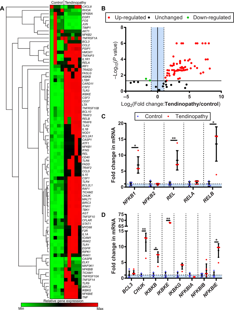
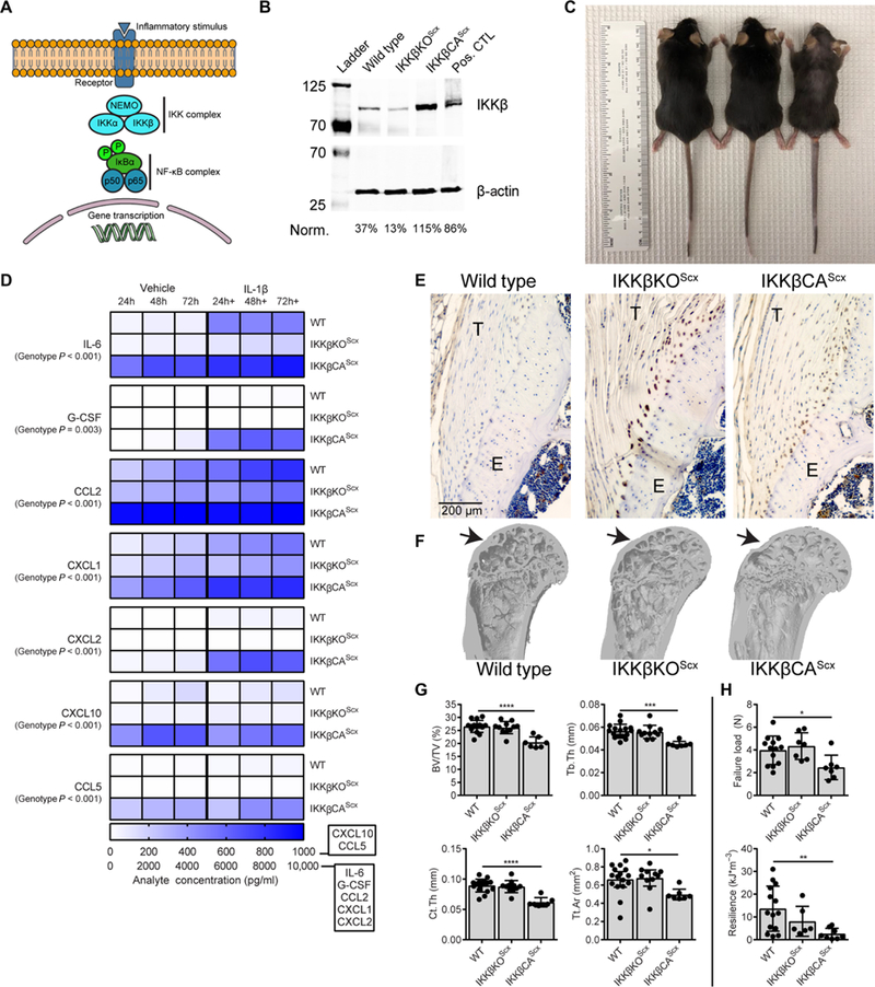
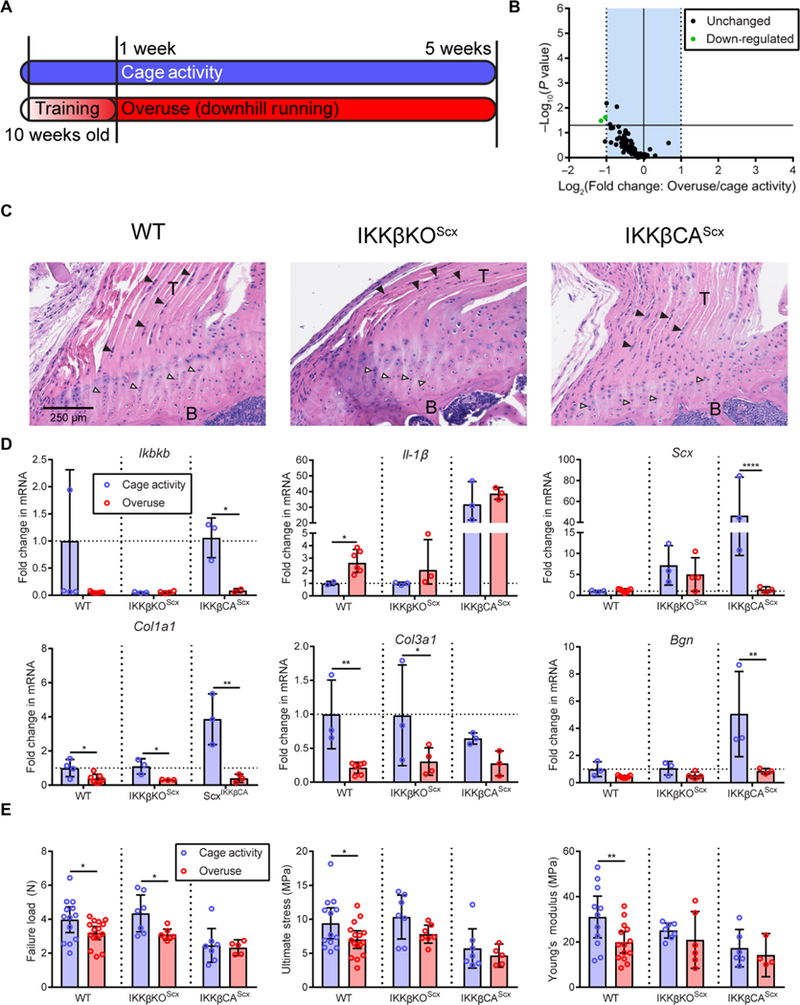
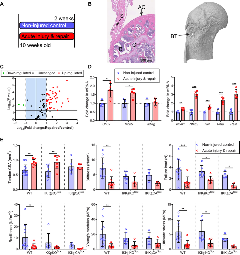
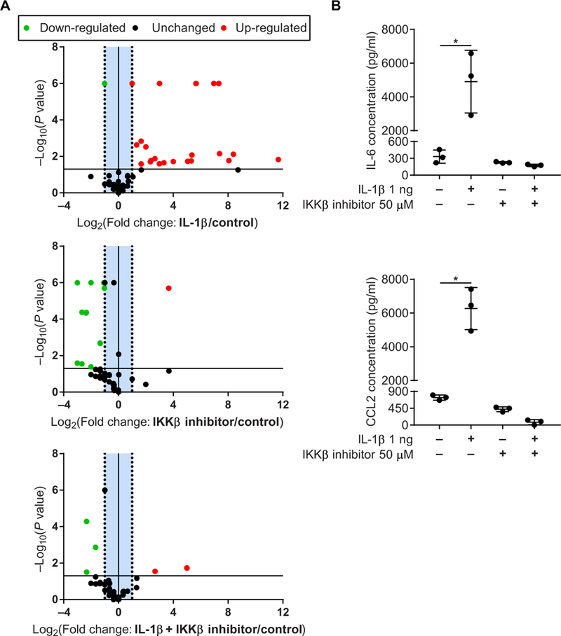
Tendon disorders represent the most common musculoskeletal complaint for which patients seek medical attention; inflammation drives tendon degeneration before tearing and impairs healing after repair. Clinical evidence has implicated the nuclear factor kappa-light-chain-enhancer of activated B cells (NF-κB) pathway as a correlate of pain-free return to function after surgical repair. However, it is currently unknown whether this response is a reaction to or a driver of pathology. Therefore, we aimed to understand the clinically relevant involvement of the NF-κB pathway in tendinopathy, to determine its potential causative roles in tendon degeneration, and to test its potential as a therapeutic candidate. Transcriptional profiling of early rotator cuff tendinopathy identified increases in NF-κB signaling, including increased expression of the regulatory serine kinase subunit IKKβ, which plays an essential role in inflammation. Using cre-mediated overexpression of IKKβ in tendon fibroblasts, we observed degeneration of mouse rotator cuff tendons and the adjacent humeral head. These changes were associated with increases in proinflammatory cytokines and innate immune cells within the joint. Conversely, genetic deletion of IKKβ in tendon fibroblasts partially protected mice from chronic overuse-induced tendinopathy. Furthermore, conditional knockout of IKKβ improved outcomes after surgical repair, whereas overexpression impaired tendon healing. Accordingly, targeting of the IKKβ/NF-κB pathway in tendon stromal cells may offer previously unidentified therapeutic approaches in the management of human tendon disorders.


Figures

Fig. 1.
Fig. 1.. NF-κB signaling in clinical tendinopathy.
(A) Heat map of NF-κB profiling gene expression array from healthy human hamstrings tendons (control, n = 4) and early-stage diseased tendons (tendinopathy, n = 5). (B) Volcano plot of NF-κB profiling gene expression array. P < 10−6 are scaled for visualization purposes. (C) NF-κB complex protein coding genes NFKB1, REL, and RELB in control and tendinopathy tendon samples. (D) Regulatory NF-κB protein coding genes CHUK, IKBKB, IKBKE, and NFKBIE in control and tendinopathy tendon samples. Data are shown as means ± SD with individual points representing biologically independent samples. Statistically significant differences were calculated using multiple t tests with Holm-Šídák correction, **P < 0.01 and *P < 0.05.
Fig. 2.
Fig. 2.. Modulation of IKKβ expression in murine tendon fibroblasts.
(A) Schematic of NF-κB signaling and gene transcription. NF-κB signaling was controlled by targeting inhibitor of NF-κB kinase subunit p (IKKβ), which acts upstream of the NF-κB complex. Tendon fibroblast IKKβ modulation was achieved by deletion of IKKβ (IKKβKOScx) and activation of IKKβ (IKKβCAScx) using Cre-loxP-mediated recombination under the Scx promoter. (B) Expression of IKKβ in tendon fibroblasts from WT, IKKβKOScx, and IKKβCAScx mice. Cultured mouse osteoclasts were used as a positive control (pos. CTL) (51). (C) Photograph of 16-week-old mice to demonstrate hair loss. (D) Secreted cytokines and growth factors in vehicle and IL-1β-treated tendon fibroblasts from WT, IKKβKOScx, and IKKβCAScx mice (n = 5 per group). (E) Immunolabeling for CD68 (brown) in the supraspinatus tendon from WT, IKKβKOScx,and IKKβCAScx mice. T, tendon; E, enthesis. (F) Microcomputed tomography (μCT) three-dimensional reconstruction of coronal section from proximal humerus. Arrows denote the supraspinatus tendon attachment site. (G) Quantification of bone morphometry: Bone volume normalized to total volume (BV/TV), trabecular thickness (Tb.Th), cortical thickness (Ct.Th), and total cortical area (Tt.Ar) (n = 8 to 9 per genotype). (H) Quantification of mechanical properties of the supraspinatus tendon-to-bone attachment (n = 8 to 9 per genotype). Data are shown as means ± SD with individual points representing biologically independent samples. Statistically significant differences were calculated using one-way analysis of variance (ANOVA) (genotype) with Fisher’s least significant difference (LSD) post hoc test. ****P < 0.0001, ***P < 0.001, **P < 0.01, *P < 0.05.
Fig. 3.
Fig. 3.. Modulation of IKK|β/NF-kB signaling with chronic overuse.
(A) Ten-week-old mice were subjected to a chronic overuse protocol with 1 week of progressive training, followed by 4 weeks of downhill running. Control mice were permitted normal cage activity. (B) NF-κB pathway-related gene regulation due to overuse. (C) Hematoxylin and eosin (H&E) images of WT, IKKβKOScx, and IKKβCAScx mice. B, bone; black arrowhead, spindle-shaped tendon fibroblast; white arrowhead, enthesis chondrocyte. (D) mRNA expression of Ikbkb, IL-1β, Scx, Col1a1, Col3a1, and Bgn in tendon from control cage-active or treadmill overuse-subjected WT (n = 4 to 6 per group), IKKβKOScx (n = 3 to 4 per group), and IKKβCAScx (n = 3 per group) mice. (E) Failure load, ultimate stress, and Young’s modulus of the supraspinatus tendon-to-bone attachment in cage-active and treadmill overuse-subjected mice (n = 5 to 13 per group). Data are shown as means ± SD with individual points representing biologically independent samples. Statistically significant differences were calculated using two-way ANOVA (genotype, overuse) with Fisher’s LSD post hoc test. **P < 0.01 and *P < 0.05.
Fig. 4.
Fig. 4.. Modulation of IKKβ/NF-κB signaling with acute supraspinatus injury and repair.
(A) Experimental protocol. Ten-week-old mice were subjected to a unilateral acute injury of the supraspinatus tendon and immediate repair, followed by 2 weeks of recovery. Sham operations were performed on contralateral limbs. (B) H&E image ofthe repaired tendon and new bone formation around the suture tunnel after 2 weeks of recovery. GP, growth plate; S, suture hole; AC, articular cartilage. The μCT image (right) shows the bone tunnel (BT) below the epiphysis. (C) NF-κB signaling gene expression 2 weeks after acute injury and repair (n = 4 to 5 per group). (D) mRNA expression of IKK complex-related genes and NF-κB complex-related genes in tendon 2 weeks after recovery (n = 4 to 5 per group). (E) Quantification of murine tendon cross-sectional area (CSA), stiffness, failure load, resilience, Young’s modulus, and ultimate stress 2 weeks after injury and repair (n = 4 to 5 per group). Data are shown as means ± SD with individual points representing biologically independent samples. Statistically significant differences were calculated using two-way ANOVA (genotype, injury) with Fisher’s LSD post hoc test. ***P < 0.01, **P <0.01,*P <0.05.
Fig. 5.
Fig. 5.. Small-molecule inhibition of IKKβ in an in vitro model of inflammation.
(A) Volcano plots of NF-κB signaling array in healthy human tendon fibroblasts treated with IL-1β with or without IKKβ inhibitor. (B) Proinflammatory cytokines IL-6 and CCL-2 produced by healthy human tendon fibroblasts treated with IL-1β with or without IKKβ inhibitor. Data are shown as means ± SD. Statistically significant differences were calculated using one-way ANOVA (treatment) with Fisher’s LSD post hoc test. *P < 0.05.

Comment in

  • Targeting NF-κB in tendinopathy.
    McHugh J. McHugh J. Nat Rev Rheumatol. 2019 May;15(5):251. doi: 10.1038/s41584-019-0206-x. Nat Rev Rheumatol. 2019. PMID: 30914773 No abstract available.

References

    1. Jordan KP, Jöud A, Bergknut C, Croft P, Edwards JJ, Peat G, Petersson IF, Turkiewicz A, Wilkie R, Englund M, International comparisons of the consultation prevalence of musculoskeletal conditions using population-based healthcare data from England and Sweden. Ann. Rheum. Dis. 73, 212–218 (2014). - PMC - PubMed
    1. Riley G, Tendinopathy—From basic science to treatment. Nat. Clin. Pract. Rheumatol. 4, 82–89 (2008). - PubMed
    1. Mather RC III, Koenig L, Acevedo D, Dall TM, Gallo P, Romeo A, Tongue J, Williams G Jr., The societal and economic value of rotator cuff repair. J. Bone Joint Surg. Am. 95, 1993–2000 (2013). - PMC - PubMed
    1. Beard DJ, Rees JL, Cook JA, Rombach I, Cooper C, Merritt N, Shirkey BA, Donovan JL, Gwilym S, Savulescu J, Moser J, Gray A, Jepson M, Tracey I, Judge A, Wartolowska K, Carr AJ, Ahrens P, Baldwick C, Brinsden M, Brownlow H, Burton D, Butt MS, Carr A, Charalambous CP, Conboy V, Dennell L, Donaldson O, Drew S, Dwyer A, Gidden D, Hallam P, Kalogrianitis S, Kelly C, Kulkarni R, Matthews T, McBirnie J, Patel V, Peach C, Roberts C, Robinson D, Rosell P, Rossouw D, Senior C, Singh B, Sjolin S, Taylor G, Venkateswaran B, Woods D, Arthroscopic subacromial decompression for subacromial shoulder pain (CSAW): A multicentre, pragmatic, parallel group, placebo-controlled, three-group, randomised surgical trial. Lancet 391, 329–338 (2018). - PMC - PubMed
    1. Ketola S, Lehtinen JT, Arnala I, Arthroscopic decompression not recommended in the treatment of rotator cuff tendinopathy: A final review of a randomised controlled trial at a minimum follow-up often years. Bone Joint J. 99-B, 799–805 (2017). - PubMed

Publication types

LinkOut - more resources