. 2019 Feb 27;10(1):959.
doi: 10.1038/s41467-019-08895-7.

Circulating myocardial microRNAs from infarcted hearts are carried in exosomes and mobilise bone marrow progenitor cells

Affiliations

Circulating myocardial microRNAs from infarcted hearts are carried in exosomes and mobilise bone marrow progenitor cells

Min Cheng et al. Nat Commun. .

Abstract

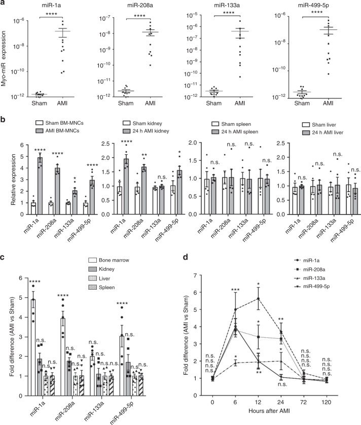
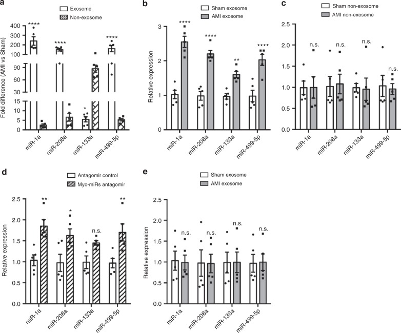
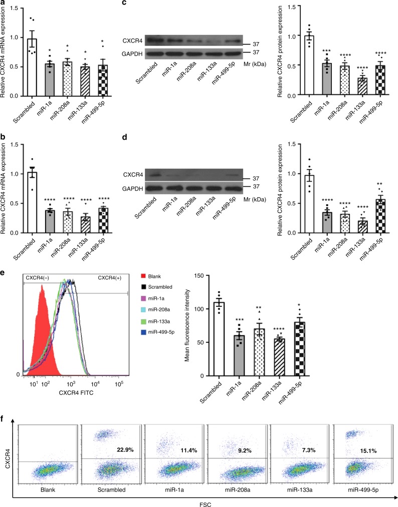
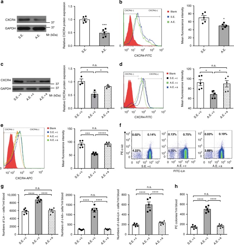
Myocardial microRNAs (myo-miRs) are released into the circulation after acute myocardial infarction (AMI). How they impact remote organs is however largely unknown. Here we show that circulating myo-miRs are carried in exosomes and mediate functional crosstalk between the ischemic heart and the bone marrow (BM). In mice, we find that AMI is accompanied by an increase in circulating levels of myo-miRs, with miR-1, 208, and 499 predominantly in circulating exosomes and miR-133 in the non-exosomal component. Myo-miRs are imported selectively to peripheral organs and preferentially to the BM. Exosomes mediate the transfer of myo-miRs to BM mononuclear cells (MNCs), where myo-miRs downregulate CXCR4 expression. Injection of exosomes isolated from AMI mice into wild-type mice downregulates CXCR4 expression in BM-MNCs and increases the number of circulating progenitor cells. Thus, we propose that myo-miRs carried in circulating exosomes allow a systemic response to cardiac injury that may be leveraged for cardiac repair.


Conflict of interest statement

The authors declare no competing interests.

Figures

Fig. 1
Fig. 1
Myo-miRs are released into PB following AMI and transported into BM-MNCs. AMI and Sham surgeries were performed in C57BL/6 mice; then at various time points, the plasma, BM-MNCs, and different organs were isolated and subjected to qRT-PCR analyzes of myo-miRs, miR-1a, 208a, 133a, and 499-5p. a myo-miR levels in the plasma 6 h post-surgery (n = 10 animals per group). b myo-miR levels in the BM-MNCs, kidney, spleen, and liver 12 h post-surgeries, expressed relative to the levels in the intact controls (n = 5 animals per group), and c the fold difference in AMI vs. Sham mice (n = 5 animals per group). *p < 0.05, **p < 0.01, ***p < 0.001, ****p < 0.0001 vs. Sham; n.s., no significant. d The fold difference of myo-miR levels in the BM-MNCs of AMI vs. Sham mice at 0, 6, 12, 24, 72, and 120 h post-surgery. *p < 0.05 **p < 0.01, ***p < 0.001 vs. Sham at same time point. n = 5 animals per group per time point. An unpaired t test was used in a and a two-way ANOVA was used in b, c, and d for statistical analysis. Error bars represent mean ± s.e.m
Fig. 2
Fig. 2
Exosomes mediate the transfer of circulating myo-miRs into BM-MNCs. Exosomes and the non-exosomal component were isolated from the plasma of AMI and Sham mice 6 h post-surgery. a The levels of myo-miRs in the exosomes and the non-exosomal component were analyzed by qRT-PCR and expressed as fold difference in AMI vs. Sham mice. *p < 0.05, ****p < 0.0001 vs. Sham. n = 5 animals per group. b, c In vitro, freshly-isolated mouse BM-MNCs (2 × 107/well) were treated for 12 h with the exosomes (20 μg) (b) or non-exosomal component (150 μl) (c) from AMI or Sham mice; then, the levels of myo-miRs in the BM-MNCs were quantified with qRT-PCR. **p < 0.01, ****p < 0.0001 vs. treatment with Sham exosomes or Sham non-exosomal component; n.s. not significant. n = 5 biologically independent samples per group. d, e In vivo, exosomes (40 μg in 300 μL PBS/mouse) (d) or the non-exosomal component (300ul/mouse) (e) from AMI or Sham mice was i.v. injected into intact C57BL/6 mice; 12 h later, the levels of myo-miRs in the recipient BM-MNCs were quantified via qRT-PCR. *p < 0.05, **p < 0.01 vs. treatment with Sham exosomes or Sham non-exosomal component; n.s., not significant. n = 5 animals per group. A two-way ANOVA was used for statistical analysis. Error bars represent mean ± s.e.m
Fig. 3
Fig. 3
Myo-miRs downregulate CXCR4 expression in BM-MNCs and MSCs in vitro. BM-MNCs (2 × 106/well) and MSCs (0.5 × 106/well) were transfected with synthesized miR-1a, miR-208a, miR-133a, or miR-499-5p mimics or with a scrambled miR (final concentrations: 50 nM); 24 h later, CXCR4 mRNA expression in the BM-MNCs (a, c, e) and MSC (b, d, f) was quantified by RT-PCR (a, b), Western-blotting (c, d), and flow cytometry (e, f). *p < 0.05, **p < 0.01, ***p < 0.001, ****p < 0.0001 vs. Scrambled. n = 5 biologically independent samples per group. A one-way ANOVA was used for statistical analysis. Error bars represent mean ± s.e.m
Fig. 4
Fig. 4
Exosomal transfer of myo-miRs downregulates CXCR4 in BM-MNCs and induces BM PC mobilization. Exosomes were isolated from the plasma of AMI mice (AMI-exosomes, A.E.) or Sham mice (Sham-exosomes, S.E.) 6 h post-surgery. ad In vitro, BM-MNCs (2 × 107/well) were cultured for 24 h with A.E. (20ug), S.E. (20 μg), A.E. plus 10 nM non-targeting scrambled control sequence (c) (A.E. + c), S.E. + c, or A.E. plus 10 nM (4 × 2.5 nM each) antagomir (A.E. + a), then quantified for CXCR4 expression by Western-blotting (a, c; n = 5 biologically independent samples per group for a, n = 3 biologically independent samples per group for c) and flow-cytometry (b, d, n = 5 biologically independent samples per group). *p < 0.05, ***p < 0.001. eh In vivo, C57BL/6 mice were injected with a (80 mg [20 mg each] antagomir/kg body weight/day) or c (80 mg/kg/day) for 3 consecutive days, then subjected to AMI or Sham surgery and 6 h later, to isolation of plasma exosomes. The isolated exosomes were subsequently i.v. injected into intact C57BL/6 mice (40 μg diluted in 300 μL PBS/mouse) and 12 h later, BM-MNCS and PB-MNCs were isolated from the recipient mice for assessments of PC mobilization. eg Flow cytometry analyzes for CXCR4 expression in BM-MNCs (e), percentages of c-kit+, Lin, and c-kit+Lin cells in the PB-MNCs (f), and calculation of absolute c-kit+, Lin, and c-kit+Lin cell numbers per 1 ml PB (g). h The colony-forming PCs in the PB-MNCs were evaluated via colony formation assay. ***p < 0.001, ****p < 0.0001, n.s., no significant. n = 5 animals per group. An unpaired t test was used in (a, b) and a one-way ANOVA was used in (ch) for statistical analysis. Error bars represent mean ± s.e.m

References

    1. Losordo DW, Vaughan DE. Going mobile: enhanced recovery from myocardial infarction via stem cell mobilization and homing for tissue repair. J. Am. Coll. Cardiol. 2014;63:2873–2874. doi: 10.1016/j.jacc.2014.02.571. - DOI - PubMed
    1. Afzal MR, et al. Adult bone marrow cell therapy for ischemic heart disease: evidence and insights from randomized controlled trials. Circ. Res. 2015;117:558–575. doi: 10.1161/CIRCRESAHA.114.304792. - DOI - PMC - PubMed
    1. Jonas S, Izaurralde E. Towards a molecular understanding of microRNA-mediated gene silencing. Nat. Rev. Genet. 2015;16:421–443. doi: 10.1038/nrg3965. - DOI - PubMed
    1. Corsten MF, et al. Circulating microRNA-208b and microRNA-499 reflect myocardial damage in cardiovascular disease. Circ. Cardiovasc. Genet. 2010;3:499–506. doi: 10.1161/CIRCGENETICS.110.957415. - DOI - PubMed
    1. Kuwabara Y, et al. Increased microRNA-1 and microRNA-133a levels in serum of patients with cardiovascular disease indicate myocardial damage. Circ. Cardiovasc. Genet. 2011;4:446–454. doi: 10.1161/CIRCGENETICS.110.958975. - DOI - PubMed

Publication types

MeSH terms