. 2019 Feb 27;9(1):2933.
doi: 10.1038/s41598-018-37588-2.

Photoacoustic Imaging for Image-guided Endovenous Laser Ablation Procedures

Affiliations

Photoacoustic Imaging for Image-guided Endovenous Laser Ablation Procedures

Yan Yan et al. Sci Rep. .

Abstract

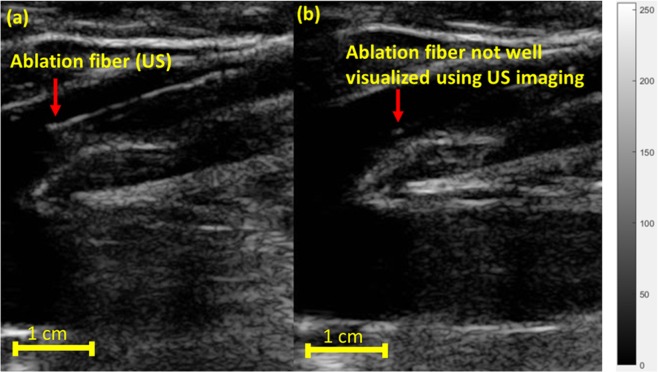
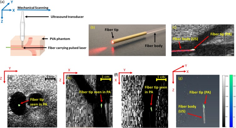
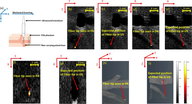
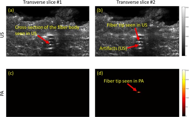
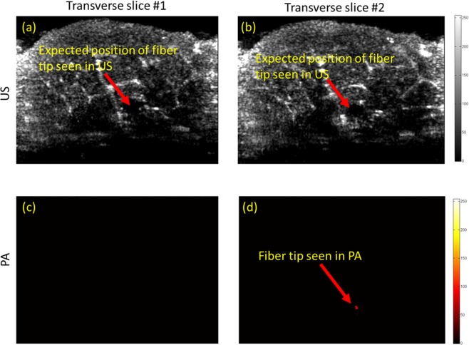
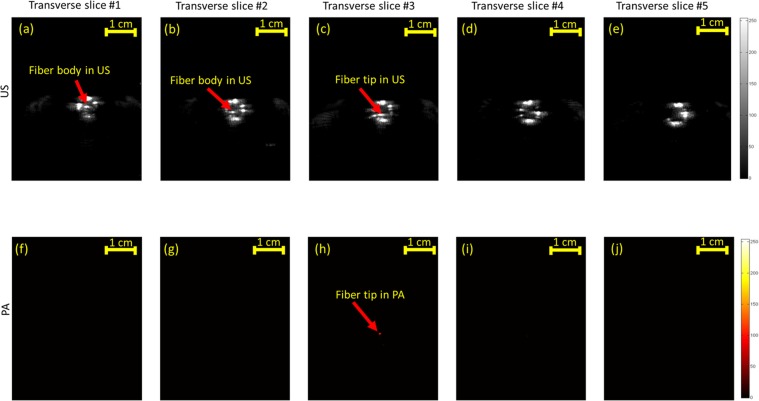
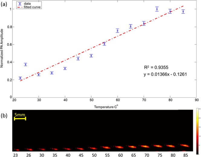
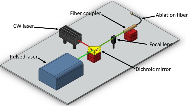
Accurate fiber tip tracking is a critical clinical problem during endovenous laser ablation (EVLA) of small perforating veins. Currently, ultrasound (US) imaging is the gold-standard modality for visualizing and for accurately placing the ablation fiber within the diseased vein. However, US imaging has limitations such as angular dependency and comet tail artifacts. In addition, EVLA is often performed without any real-time temperature monitoring, which could lead to an insufficient thermal dose or overheating the surrounding tissue. We propose a new technique that combines US and photoacoustic (PA) imaging for concurrent ablation fiber tip tracking and real-time temperature monitoring during EVLA procedures. Our intended implementation of PA imaging for fiber tracking requires minimal modification of existing systems, which makes this technology easy to adopt. Combining US and PA imaging modalities allows for simultaneous visualization of background anatomical structures as well as high contrast, artifact-free, and angle-independent localization of the ablation fiber tip. Preliminary data demonstrates that changes in the amplitude of the PA signal can be used to monitor the localized temperature at the tip of the ablation fiber, which will be invaluable during EVLA procedures. These improvements can enhance the physician's accuracy in performing EVLA procedures and will have a significant impact on the treatment outcomes.


Conflict of interest statement

The authors declare no competing interests.

Figures

Figure 1
Figure 1
Sagittal US images visualizing the ablation fiber in a diseased vein during EVLA. (a) US imaging visualizes the ablation fiber inside the diseased vein. (b) Due to angular dependency and improper alignment of the US transducer with respect to the ablation fiber, it is not clearly visualized. Images were obtained from Henry Ford hospital (Detroit, Michigan).
Figure 2
Figure 2
(a) Experimental setup for straight fiber tip tracking. (b) image showing the ablation fiber used, the ablation fiber has a gold covering at its tip. (c) Fiber tip seen using PA imaging, body of the fiber seen using US imaging. Appearance of the straight fiber in combined US and thresholded PA imaging in (d) transverse, (e) sagittal, and (f) coronal planes. (g) Volumetric image of the fiber in both US and thresholded PA images indicating that PA is only visualizing the fiber tip while US imaging shows the whole fiber body. Supplementary Movie 1 shows the volumetric US and PA of the phantom with straight fiber.
Figure 3
Figure 3
(a) Experimental setup for angled fiber tip tracking. US imaging of the angled fiber indicating the pulse-echo US limitations in tracking the angled fiber in (c,e,g,i). Thresholded PA imaging of the angled fiber inside vessel-mimicking phantom, superimposed over B-mode US image of the phantom in (b,d,f,h). Supplementary Movies 2 and 3 show the volumetric US and PA of the phantom with angled fiber.
Figure 4
Figure 4
US (a,b) and PA (c,d) images of a fiber inside a porcine tissue in straight orientation. US imaging is unable to track the fiber inside the porcine tissue in (a,b). The accurate location of the fiber tip is clearly seen using thresholded PA imaging inside the porcine meat tissue in (c,d).
Figure 5
Figure 5
US (a,b) and PA (c,d) images of a fiber inside a porcine tissue at an angle of 30 degrees. Due to angular dependency issues, US imaging is unable to track the fiber inside the porcine tissue in (a,b). The accurate location of the fiber tip is clearly seen using thresholded PA imaging inside the porcine tissue in (c,d).
Figure 6
Figure 6
US (ae) and PA (fj) mechanically scanned transverse images of a fiber tip placed inside a blood-filled tube at behind (panels a–b and f–g) of the tip, at the fiber tip (panels c,h) and after passing the fiber (panels d–e, and i–j). US imaging is unable to distinguish between the fiber tip and the cross section of the fiber in (ac). The accurate location of the fiber tip is clearly seen using thresholded PA imaging in (h).
Figure 7
Figure 7
(a) PA imaging to monitor the changes in amplitude of the PA signal with increase in the surrounding temperature. (a) Plot indicating normalized amplitude of the PA signal obtained versus surrounding temperature, blue dots indicate measurements and the red dashed line represents the linear fit. (b) PA images of the fiber tip at different surrounding temperature. The increase in PA amplitude as well as the effect of speed of sound variation can be clearly seen in (b).
Figure 8
Figure 8
Block diagram of the combined PA and EVLA system. The ablation fiber carries the combined beam from the pulse laser and the CW ablation laser through a dichroic mirror and provides simultaneous ablation fiber tip tracking and real time temperature monitoring inside the vein. Supplementary Movie 4 shows the combined CW laser beam (λ = 808 nm) and pulsed laser beam (λ = 532 nm) using proposed optics.

References

    1. Informed Health Online. Institute for Quality and Efficiency in Health Care (IQWiG): Cologne, Germany (2016).
    1. Nijsten T, et al. Minimally invasive techniques in the treatment of saphenous varicose veins. J. Am. Acad. Dermatol. 2009;60:110–119. doi: 10.1016/j.jaad.2008.07.046. - DOI - PubMed
    1. Jones, R. H. & Carek, P. J. Management of varicose veins. Am. Fam. Physician78 (2008). - PubMed
    1. Subramonia S, Lees T. The treatment of varicose veins. The Annals The Royal Coll. Surg. Engl. 2007;89:96–100. - PMC - PubMed
    1. Ramelet A-A. Phlebectomy. Int. Angiol. 2002;21:46. - PubMed

Publication types