Observational Study
. 2019 Feb 27;9(1):2874.
doi: 10.1038/s41598-019-39726-w.

Neutrophil Dysfunction in the Airways of Children with Acute Respiratory Failure Due to Lower Respiratory Tract Viral and Bacterial Coinfections

Affiliations
Observational Study

Neutrophil Dysfunction in the Airways of Children with Acute Respiratory Failure Due to Lower Respiratory Tract Viral and Bacterial Coinfections

Jocelyn R Grunwell et al. Sci Rep. .

Abstract

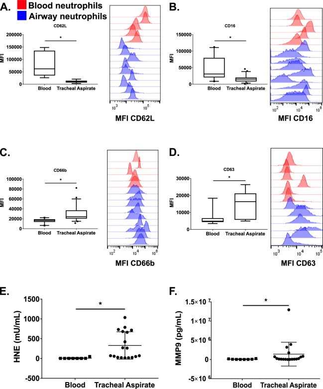
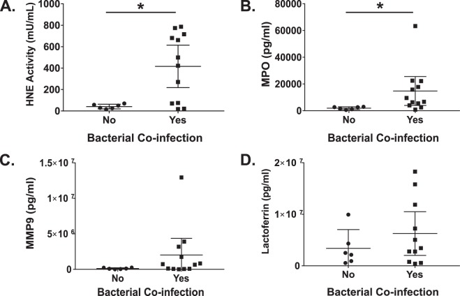
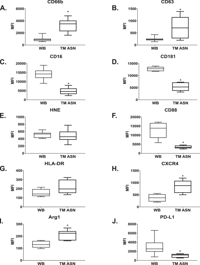
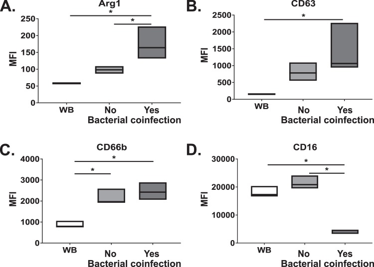
Neutrophils are recruited to the airways of patients with acute respiratory distress syndrome (ARDS) where they acquire an activated pro-survival phenotype with an enhanced respiratory burst thought to contribute to ARDS pathophysiology. Our in vitro model enables blood neutrophil transepithelial migration into cell-free tracheal aspirate fluid from patients to recapitulate the primary airway neutrophil phenotype observed in vivo. Neutrophils transmigrated through our model toward airway fluid from children with lower respiratory viral infections coinfected with bacteria had elevated levels of neutrophil activation markers but paradoxically exhibited an inability to kill bacteria and a defective respiratory burst compared with children without bacterial coinfection. The airway fluid from children with bacterial coinfections had higher levels of neutrophil elastase activity, as well as myeloperoxidase levels compared to children without bacterial coinfection. Neutrophils transmigrated into the aspirate fluid from children with bacterial coinfection showed decreased respiratory burst and killing activity against H. influenzae and S. aureus compared to those transmigrated into the aspirate fluid from children without bacterial coinfection. Use of a novel transmigration model recapitulates this pathological phenotype in vitro that would otherwise be impossible in a patient, opening avenues for future mechanistic and therapeutic research.


Conflict of interest statement

Drs. Grunwell, Fitzpatrick, and Tirouvanziam received support for research from the National Institutes of Health. None of the authors have any conflicts of interest. Dr. Grunwell is supported by the Atlanta Pediatric Scholars Program grant K12HD072245.

Figures

Figure 1
Figure 1
Characterization of blood and airway neutrophils markers by flow cytometry and markers of neutrophil activation in the plasma and airway fluid. Box plots of neutrophil cell surface markers for (A) CD62L, (B) CD16, (C) CD66b, and (D) CD63 in the blood (n = 8) and airway (n = 18) collected within 24 hours of endotracheal intubation (Day 1). Examples of histograms of the primary flow cytometry data are shown beside the box plot for each marker. Red and blue histograms represent blood and airway neutrophils, respectively. (E) Human neutrophil elastase activity assay and (F) matrix metalloproteinase 9 (MMP-9) protein levels from Day 1 plasma and cell-free airway fluid from tracheal aspirate samples. Box plots depict median values, the box edges are the 25th to 75th interquartile ranges (IQR), and the whiskers are the 5−95% confidence intervals. *p < 0.05.
Figure 2
Figure 2
Characterization of blood (n = 8) and airway (n = 18) neutrophil cell surface markers of activation by flow cytometry. Samples were collected within 24 hours of endotracheal intubation (Day 1). Surface expression of integrins (A) CD11b, (B) CD11c, (C) CD49d, and (D) CD54, the immunoglobulin G receptor (E) CD32, (F) MHC II receptor, HLA-DR, and complement receptors (G) CD35 (C3b/C4b, CR1), and (H) CD88 (C5aR), and of the IL8 receptors (I) CD181 and (J) CD182. Box plots depict median values, the box edges are the 25th to 75th interquartile ranges (IQR), and the whiskers are the 5−95% confidence intervals. *p < 0.05.
Figure 3
Figure 3
Airway supernatant (ASN) markers of neutrophils activation analyzed by the absence (n = 9–11) or presence (n = 19–21) of a respiratory bacterial co-infection from tracheal aspirate samples obtained within 24 hours of endotracheal intubation (Day 1 samples). (A) Human neutrophil elastase (HNE) activity and (B) myeloperoxidase levels are elevated in children with a respiratory bacterial co-infection compared to those without a bacterial co-infection. There is no significant difference in (C) matrix metalloproteinase 9 (MMP-9) or (D) lactoferrin protein levels in children with a respiratory bacterial co-infection compared to those without a bacterial co-infection. The central line is the mean value with whiskers representing the standard deviation. Samples were compared using the Mann-Whitney U test. *p < 0.05.
Figure 4
Figure 4
Characterization of cell surface markers of activation by flow cytometry of donor neutrophils from whole blood (WB) (n = 6) and transmigrated through the small-airways model for 14 hours towards airway supernatant (TM ASN) (n = 6–9). (A) CD66b, (B) CD63, (C) CD16, (D) CD181, (E) HNE, (F) CD88, (G) HLA-DR, (H) CXCR4 (CD184), (I) Arg-1, (J) PD-L1. Box plots depict median values, the box edges are the 25th to 75th interquartile ranges (IQR), and the whiskers are the 5−95% confidence intervals. Samples were compared using the Mann-Whitney U test. *p < 0.05.
Figure 5
Figure 5
Transmigration through the airway model primes healthy donor neutrophils to activate the respiratory burst when stimulated by N-formylmethionyl-leucine-phenylalanine (fMLF) as measured by dihydrorhodamine (DHR) flow cytometry assay. (A) Representative flow cytometry histograms depicting healthy donor neutrophil DHR response to fMLF without (blue histogram) and with priming by GM-CSF (100 ng/mL; green histogram) for 30 minutes at 37 °C, 5% CO2. PMA-stimulated (100 pg/mL) neutrophils serve as a positive control (orange histogram). Unstimulated, DHR loaded neutrophils are shown as a negation control (red histogram). (B) Representative flow cytometry data depicting the DHR fluorescence to fMLF stimulus for transmigrated to LTB4 (red histograms), transmigrated to pooled Day 1 ASN (blue histograms) and neutrophils co-incubated with pooled Day 1 ASN (green histograms) for one donor. PMA (magenta histogram) is included as a maximal respiratory burst positive control. (C) Change in the DHR response to fMLF stimulation for neutrophils allowed to transmigrate for 14 hours toward LTB4, ASN with no bacterial coinfection (low HNE), or ASN with bacterial coinfection (high HNE) diluted 1:3 in serum-free media. Data are reported as the mean and standard deviation for three donors and analyzed using ANOVA with a post-hoc Tukey test for multiple comparisons. *p < 0.05. (D) Representative flow cytometry histogram from one neutrophil donor transmigrated as described in C).
Figure 6
Figure 6
Box plots depict surface marker changes for neutrophils from 3 donors transmigrated as in C): (A) Arg1, (B) CD63, C) CD66b, and (D) CD16. Box plots depict median values, the box edges are the minimum and maximum values. *p < 0.05.
Figure 7
Figure 7
Percent bacterial killing of (A) Haemophilus influenzae or (B) Staphylococcus aureus by neutrophils from 3 donors allowed to transmigrate to LTB4, or ASN with no bacterial coinfection (low HNE) or ASN with bacterial coinfection (high HNE) for 14 hours. Samples were compared using ANOVA with a post-hoc Tukey test for multiple comparisons. *p < 0.05.

References

    1. Ashbaugh DG, Bigelow DB, Petty TL, Levine BE. Acute respiratory distress in adults. Lancet. 1967;2:319–323. doi: 10.1016/S0140-6736(67)90168-7. - DOI - PubMed
    1. Khemani RG, Smith LS, Zimmerman JJ, Erickson S ; Pediatric Acute Lung Injury Consensus Conference Group. Pediatric acute respiratory distress syndrome: definition, incidence, and epidemiology: proceedings from the Pediatric Acute Lung Injury Consensus Conference. Pediatr Crit Care Med. 2015;16:S23–40. doi: 10.1097/PCC.0000000000000432. - DOI - PubMed
    1. Grommes J, Soehnlein O. Contribution of neutrophils to acute lung injury. Mol Med. 2011;17:293–307. doi: 10.2119/molmed.2010.00138. - DOI - PMC - PubMed
    1. Weiland JE, et al. Lung neutrophils in the adult respiratory distress syndrome. Clinical and pathophysiologic significance. Am Rev Respir Dis. 1986;133:218–225. doi: 10.1164/arrd.1986.133.2.218. - DOI - PubMed
    1. Juss JK, et al. Acute Respiratory Distress Syndrome Neutrophils Have a Distinct Phenotype and Are Resistant to Phosphoinositide 3-Kinase Inhibition. Am J Respir Crit Care Med. 2016;194:961–973. doi: 10.1164/rccm.201509-1818OC. - DOI - PMC - PubMed

Publication types

MeSH terms