Comparative Study
. 2019 Feb 27;9(1):2913.
doi: 10.1038/s41598-019-39770-6.

Fibroblast origin shapes tissue homeostasis, epidermal differentiation, and drug uptake

Affiliations
Comparative Study

Fibroblast origin shapes tissue homeostasis, epidermal differentiation, and drug uptake

Christian Hausmann et al. Sci Rep. .

Abstract

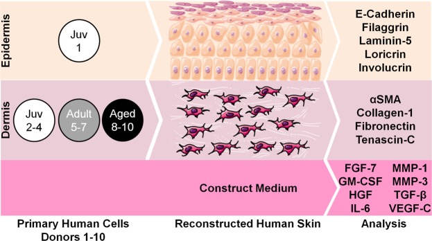
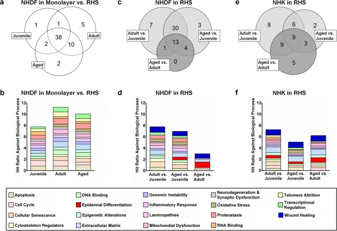
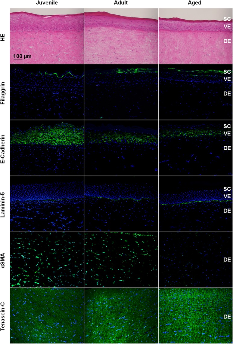
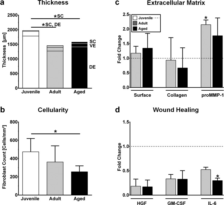
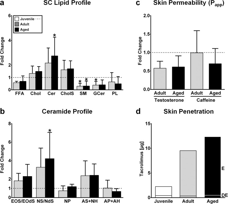
Preclinical studies frequently lack predictive value for human conditions. Human cell-based disease models that reflect patient heterogeneity may reduce the high failure rates of preclinical research. Herein, we investigated the impact of primary cell age and body region on skin homeostasis, epidermal differentiation, and drug uptake. Fibroblasts derived from the breast skin of female 20- to 30-year-olds or 60- to 70-year-olds and fibroblasts from juvenile foreskin (<10 years old) were compared in cell monolayers and in reconstructed human skin (RHS). RHS containing aged fibroblasts differed from its juvenile and adult counterparts, especially in terms of the dermal extracellular matrix composition and interleukin-6 levels. The site from which the fibroblasts were derived appeared to alter fibroblast-keratinocyte crosstalk by affecting, among other things, the levels of granulocyte-macrophage colony-stimulating factor. Consequently, the epidermal expression of filaggrin and e-cadherin was increased in RHS containing breast skin fibroblasts, as were lipid levels in the stratum corneum. In conclusion, the region of the body from which fibroblasts are derived appears to affect the epidermal differentiation of RHS, while the age of the fibroblast donors determines the expression of proteins involved in wound healing. Emulating patient heterogeneity in preclinical studies might improve the treatment of age-related skin conditions.


Conflict of interest statement

The authors declare no competing interests.

Figures

Figure 1
Figure 1
Scheme of the reconstructed human skin (RHS) used in this study. The epidermis of all RHS contained juvenile normal human keratinocytes (donor 1). Either juvenile (donor 2–4), adult (donor 5–7) or aged (donor 8–10) normal human dermal fibroblasts were used for the dermal compartment of the RHS (n = 3 for each test group). Proteins and genes were analysed from the epidermis, dermis, and medium samples. Alpha smooth muscle actin, αSMA; fibroblast growth factor-7, FGF-7; granulocyte-macrophage colony-stimulating factor, GM-CSF; hepatocyte growth factor, HGF; interleukin-6, IL-6; matrixmetalloproteinase-1/3, MMP-1/3; transforming growth factor-β, TGF-β; vascular endothelial growth factor-C, VEGF-C. This image was prepared by CH using images from Servier Medical Art under Creative Commons licence 3.0 (https://creativecommons.org/licenses/by/3.0/).
Figure 2
Figure 2
Impact of normal human dermal fibroblast (NHDF) donor age, body region, and culture on gene expression. (a,c,e) Venn diagrams showing the number of genes altered due to (a) culture conditions, (c,e) donor age (dark grey), and body region of the NHDFs (light grey). (b,d,f) Hit ratios of the altered genes for different biological processes. (a,b) Comparison of gene expression in NHDF monolayers and reconstructed human skin (RHS). (c,d) Comparison of gene expression in NHDFs from the RHS dermis and (e,f) in normal human keratinocytes from the RHS epidermis. The diagrams consider fold changes in gene expression > |1.3| and Ct values ≤ 35 for the 19 groups of biological processes; the maximum proportion of altered gene expression per biological process (hit ratio) = 1 (for details of biological processes see Table S1).
Figure 3
Figure 3
Impact of normal human dermal fibroblast donor age and body region on morphology and protein expression in reconstructed human skin. Haematoxylin and eosin staining as well as immunolocalization of filaggrin, e-cadherin, laminin-5, alpha smooth muscle actin (αSMA), and tenascin-c. Stratum corneum, SC; viable epidermis, VE; dermis, DE. Pictures are representative of at least three batches; scale bar = 100 µm.
Figure 4
Figure 4
Impact of normal human dermal fibroblast (NHDF) donor age and body region on morphology and protein expression in reconstructed human skin (RHS). (a) Thickness of the RHS layers (stratum corneum, SC; viable epidermis, VE; dermis, DE). (b) NHDF counts. (c) Fold change in the surface area and collagen I content of RHS as well as the pro-matrixmetalloproteinase-1 (proMMP-1) concentration in the construct medium. (d) Fold change in the concentration of hepatocyte growth factor (HGF), granulocyte-macrophage colony-stimulating factor (GM-CSF), and interleukin-6 (IL-6) in the construct medium. Data are representative of at least three batches. Data are presented as the mean ± SD; *p ≤ 0.05 compared to juvenile RHS; dotted line, fold changes in comparison to juvenile RHS.
Figure 5
Figure 5
Impact of normal human dermal fibroblast donor age and body region on the stratum corneum (SC) lipid composition and skin barrier function and comparison to juvenile reconstructed human skin (RHS) (ac; dotted line). (a) Fold change in the SC lipid composition of RHS for free fatty acids (FFA), cholesterol (Chol), ceramides (Cer), cholesterol sulfate (CholS), sphingomyelin (SM), glucosylceramides (GCer), and phospholipids (PL). (b) Fold change in the ceramide classes. See the Supporting Information for ceramide nomenclature. (c) Mean apparent permeability coefficient (Papp) of testosterone and caffeine. (d) Penetration of tacrolimus from ointment into RHS. Epidermis, E; dermis, DE. Graphs depict data from three batches (ac) or one batch (d) and are presented as the mean ± SD; *p ≤ 0.05 compared to juvenile RHS.

References

    1. López-Otín C, Blasco MA, Partridge L, Serrano M, Kroemer G. The hallmarks of aging. Cell. 2013;153:1194–1217. doi: 10.1016/j.cell.2013.05.039. - DOI - PMC - PubMed
    1. Smietana K, Siatkowski M, Moller M. Trends in clinical success rates. Nat Rev Drug Discov. 2016;15:379–380. doi: 10.1038/nrd.2016.85. - DOI - PubMed
    1. Fabre KM, Livingston C, Tagle DA. Organs-on-chips (microphysiological systems): tools to expedite efficacy and toxicity testing in human tissue. Exp Biol Med (Maywood) 2014;239:1073–1077. doi: 10.1177/1535370214538916. - DOI - PubMed
    1. Benam KH, et al. Engineered in vitro disease models. Annu Rev Pathol. 2015;10:195–262. doi: 10.1146/annurev-pathol-012414-040418. - DOI - PubMed
    1. Blume-Peytavi U, et al. Age-associated skin conditions and diseases: current perspectives and future options. Gerontologist. 2016;56:S230–S242. doi: 10.1093/geront/gnw003. - DOI - PubMed

Publication types

MeSH terms