

Motor Recovery after Transplantation of Bone Marrow Mesenchymal Stem Cells in Rat Models of Spinal Cord Injury
- PMID: 30814821
- PMCID: PMC6388433
- DOI: 10.1159/000487069
Motor Recovery after Transplantation of Bone Marrow Mesenchymal Stem Cells in Rat Models of Spinal Cord Injury
Abstract
Background: Neuronal tissue has a limited potential to self-renew or get repaired after damage. Cell therapies using stem cells are promising approaches for the treatment of central nervous system (CNS) injuries. However, the clinical use of embryonic stem cells is limited by ethical concerns and other scientific consequences. Bone marrow mesenchymal stromal cells (BM-MSC) could represent an alternative source of stem cells for replacement therapy. Indeed, many studies have demonstrated that MSCs can give rise to neuronal cells as well as many tissue-specific cell phenotypes.
Purpose: Motor recovery by transplantation of bone marrow MSCs in rat models of spinal cord injury (SCI).
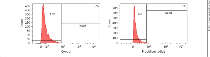
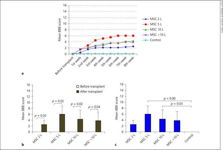
Methods: Bone marrow was collected from the femur of albino Wistar rats. MSCs were separated using the Ficoll-Paque density gradient method and cultured in Dulbecco's Modified Eagle Medium supplemented with 20% fetal bovine serum. Cultured MSC was characterized by immunohistochemistry and flow cytometry and neuronal-induced cells were further characterized for neural markers. Cultured MSCs were transplanted into the experimentally injured spinal cord of Wistar rats. Control (injured, but without cell transplantation) and transplanted rats were followed up to 8 weeks, analyzed using the Basso, Beattie, Bresnahan (BBB) scale and electromyography (EMG) for behavioral and physiological status of the injured spinal cord. Finally, the tissue was evaluated histologically.
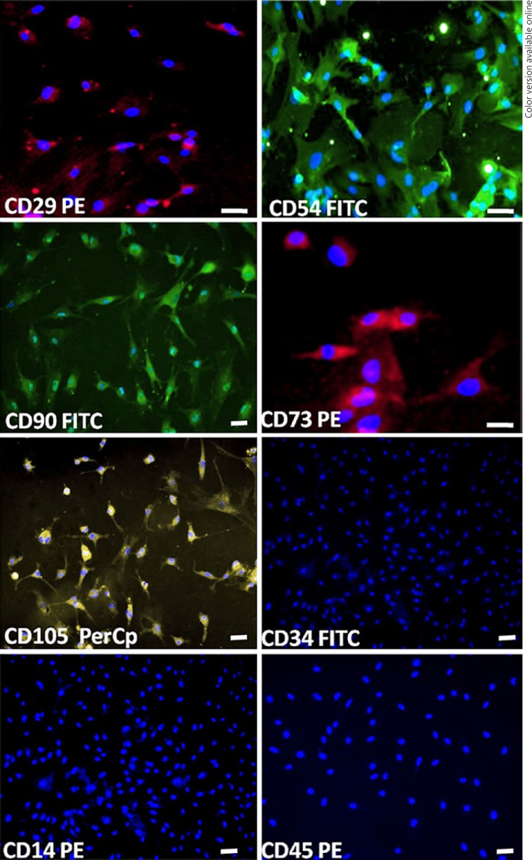
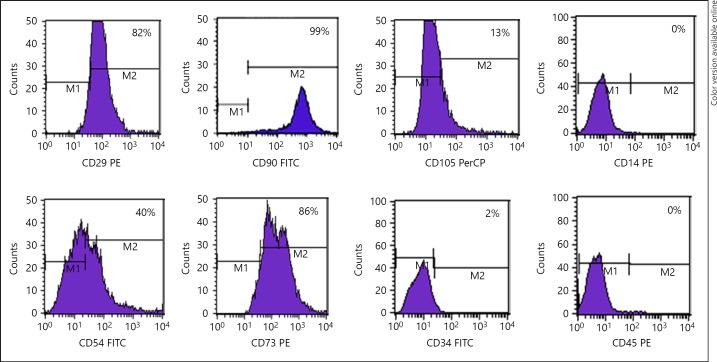
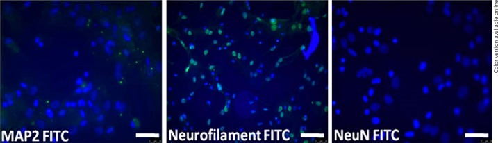
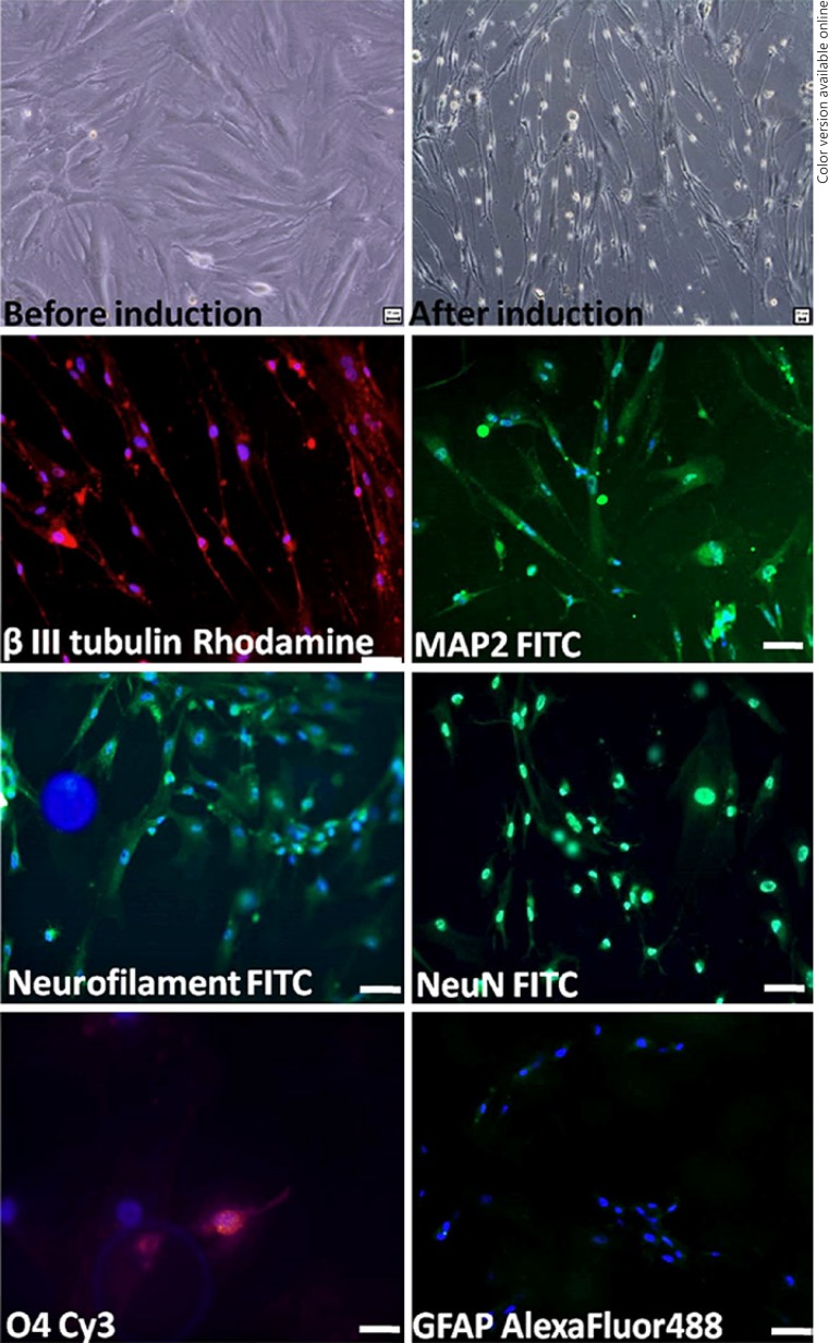
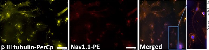
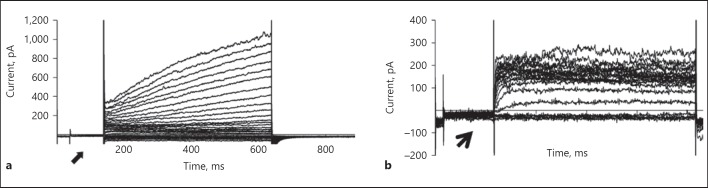
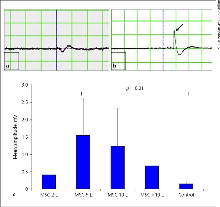
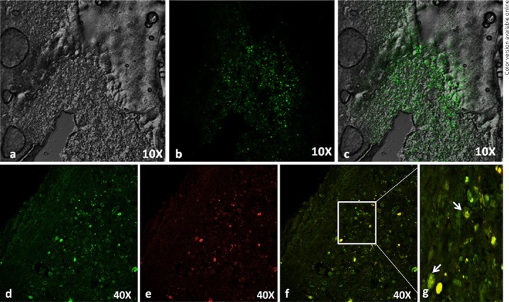
Results: Rat MSCs expressed positivity for a panel of MSC markers CD29, CD54, CD90, CD73, and CD105, and negativity for hematopoietic markers CD34, CD14, and CD45. In vitro neuronal transdifferentiated MSCs express positivity for β III tubulin, MAP2, NF, NeuN, Nav1.1, oligodendrocyte (O4), and negativity for glial fibrillary acid protein. All the treated groups show promising hind-limb motor recovery BBB score, except the control group. There was increased EMG amplitude in treated groups as compared to the control group. Green fluorescent protein (GFP)-labeled MSC survived and differentiated into neurons in the injured spinal cord, which is responsible for functional recovery.
Conclusion: Our results demonstrate that BM-MSC has the potential to repair the injured cord in rat models of SCI. Thus, BM-MSC appears to be a promising candidate for cell-based therapy in CNS injury.
Keywords: Basso, Beattie, Bresnahan score; Electromyography; Mesenchymal stem cells; Spinal cord injury; Transplantation.
Figures
References
-
- Crowe MJ, Bresnahan JC, Shuman SL, Masters JN, Beattie MS. Apoptosis and delayed degeneration after spinal cord injury in rats and monkeys. Nat Med. 1997;3:73–76. - PubMed
-
- Dumont RJ, Okonkwo DO, Verma S, Hurlbert RJ, Boulos PT, Ellegala DB, et al. Acute spinal cord injury, part I: pathophysiologic mechanisms. Clin Neuropharmacol. 2001;24:254–264. - PubMed
-
- Kwon BK, Borisoff JF, Tetzlaff W. Molecular targets for therapeutic intervention after spinal cord injury. Mol Interv. 2002;2:244–258. - PubMed
-
- Furuya T, Hashimoto M, Koda M, Okawa A, Murata A, Takahashi K, et al. Treatment of rat spinal cord injury with a Rho-kinase inhibitor and bone marrow stromal cell transplantation. Brain Res. 2009;1295:192–202. - PubMed
-
- Parr AM, Kulbatski I, Wang XH, Keating A, Tator CH. Fate of transplanted adult neural stem/progenitor cells and bone marrow-derived mesenchymal stromal cells in the injured adult rat spinal cord and impact on functional recovery. Surg Neurol. 2008;70:600–607. discussion 607. - PubMed
LinkOut - more resources
Full Text Sources
Research Materials
Miscellaneous
